
Search Count: 85
All
Selected
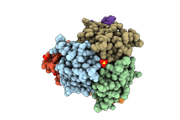 |
Organism: Homo sapiens, Squalus acanthias
Method: X-RAY DIFFRACTION Resolution:1.92 Å Release Date: 2025-11-05 Classification: Cytokine/Immune System Ligands: EDO, SO4, NI |
 |
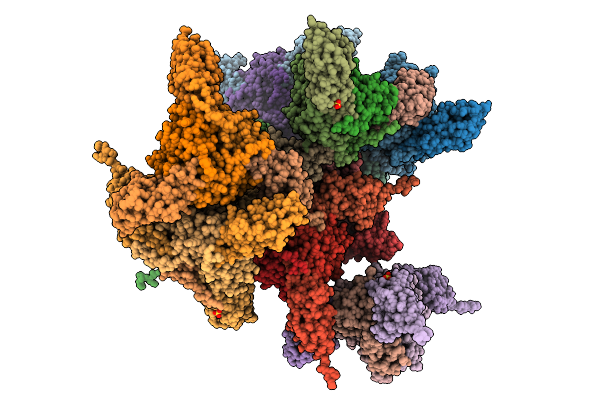
Organism: Squalus acanthias
Method: ELECTRON MICROSCOPY Release Date: 2025-10-01 Classification: MEMBRANE PROTEIN Ligands: CLR, PCW, MG, A1MA6 |
 |
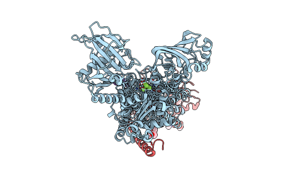
Cryo-Em Structure Of Palytoxin-Bound Na+,K+-Atpase In The Transient State Of Dephosphorylation (E2~P)
Organism: Squalus acanthias
Method: ELECTRON MICROSCOPY Release Date: 2025-10-01 Classification: MEMBRANE PROTEIN Ligands: CLR, PCW, ALF, MG, NA, A1MA6 |
 |
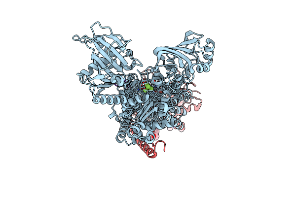
Cryo-Em Structure Of Na+,K+-Atpase That Forms A Cation Channel With Palytoxin (Atp Form)
Organism: Squalus acanthias
Method: ELECTRON MICROSCOPY Release Date: 2025-10-01 Classification: MEMBRANE PROTEIN Ligands: CLR, PCW, MG, ATP, NA, A1MA6 |
 |
Cryo-Em Structure Of Na+,K+-Atpase That Forms A Cation Channel With Palytoxin (Adp Form)
Organism: Squalus acanthias
Method: ELECTRON MICROSCOPY Release Date: 2025-10-01 Classification: MEMBRANE PROTEIN Ligands: CLR, PCW, MG, ADP, NA, A1MA6 |
 |
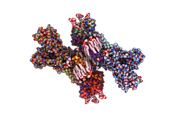
Organism: Squalus acanthias, Homo sapiens
Method: X-RAY DIFFRACTION Resolution:3.43 Å Release Date: 2025-09-10 Classification: TOXIN Ligands: GOL, SO4, CL |
 |
Organism: Squalus acanthias
Method: ELECTRON MICROSCOPY Release Date: 2023-08-09 Classification: MEMBRANE PROTEIN Ligands: CLR, PCW, MG |
 |
Organism: Squalus acanthias
Method: ELECTRON MICROSCOPY Release Date: 2022-07-13 Classification: MEMBRANE PROTEIN Ligands: K, CLR, PCW |
 |
Cryo-Em Structure Of The Na+,K+-Atpase In The E2.2K+ State After Addition Of Atp
Organism: Squalus acanthias
Method: ELECTRON MICROSCOPY Release Date: 2022-07-13 Classification: MEMBRANE PROTEIN Ligands: ATP, K, CLR, PCW |
 |
Organism: Squalus acanthias
Method: ELECTRON MICROSCOPY Release Date: 2022-04-27 Classification: MEMBRANE PROTEIN Ligands: MG, NA, ATP, CLR, PCW |
 |
Cryo-Em Structure Of Na+,K+-Atpase In The E2P State Formed By Atp In The Presence Of 40 Mm Mg2+
Organism: Squalus acanthias
Method: ELECTRON MICROSCOPY Release Date: 2022-04-27 Classification: MEMBRANE PROTEIN Ligands: MG, NA, ATP, CLR, PCW |
 |
Cryo-Em Structure Of Na+,K+-Atpase In The E2P State Formed By Inorganic Phosphate
Organism: Squalus acanthias
Method: ELECTRON MICROSCOPY Release Date: 2022-04-27 Classification: MEMBRANE PROTEIN Ligands: MG, CLR, PCW |
 |
Cryo-Em Structure Of Na+,K+-Atpase In The E2P State Formed By Atp With Istaroxime
Organism: Squalus acanthias
Method: ELECTRON MICROSCOPY Release Date: 2022-04-27 Classification: MEMBRANE PROTEIN Ligands: MG, NA, ATP, 7Q2, CLR, PCW |
 |
Cryo-Em Structure Of Na+,K+-Atpase In The E2P State Formed By Inorganic Phosphate With Istaroxime
Organism: Squalus acanthias
Method: ELECTRON MICROSCOPY Release Date: 2022-04-27 Classification: MEMBRANE PROTEIN Ligands: MG, 7Q2, CLR, PCW |
 |
Cryo-Em Structure Of Na+,K+-Atpase In The E2P State Formed By Atp With Ouabain
Organism: Squalus acanthias
Method: ELECTRON MICROSCOPY Release Date: 2022-04-27 Classification: MEMBRANE PROTEIN Ligands: MG, NA, OBN, CLR, PCW |
 |
Cryo-Em Structure Of Na+,K+-Atpase In The E2P State Formed By Inorganic Phosphate With Ouabain
Organism: Squalus acanthias
Method: ELECTRON MICROSCOPY Release Date: 2022-04-27 Classification: MEMBRANE PROTEIN Ligands: MG, OBN, CLR, PCW |
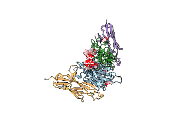 |
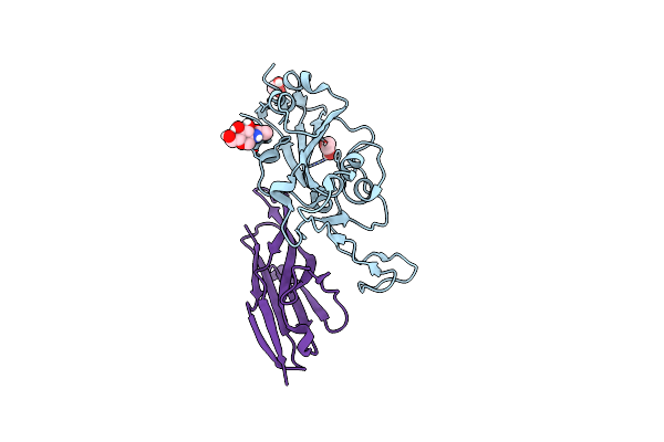
Crystal Structure Of The Sars-Cov-2 Receptor Binding Domain In Complex With Vnar 3B4
Organism: Severe acute respiratory syndrome coronavirus 2, Squalus acanthias
Method: X-RAY DIFFRACTION Resolution:1.92 Å Release Date: 2022-01-05 Classification: VIRAL PROTEIN/IMMUNE SYSTEM Ligands: EDO |
 |
Crystal Structure Of The Sars-Cov-2 Receptor Binding Domain In Complex With Vnar 2C02
Organism: Severe acute respiratory syndrome coronavirus 2, Squalus acanthias
Method: X-RAY DIFFRACTION Resolution:1.96 Å Release Date: 2022-01-05 Classification: VIRAL PROTEIN/IMMUNE SYSTEM Ligands: EDO, NAG, CL |
 |
Kinetics By X-Ray Crystallography: Tl+-Substitution Of Bound K+ In The E2.Mgf42-.2K+ Crystal After 0.75 Min.
Organism: Squalus acanthias
Method: X-RAY DIFFRACTION Resolution:2.60 Å Release Date: 2015-09-02 Classification: HYDROLASE/TRANSPORT PROTEIN Ligands: MF4, MG, K, TL, CLR, NAG |
 |
Kinetics By X-Ray Crystallography: Tl+-Substitution Of Bound K+ In The E2.Mgf42-.2K+ Crystal After 1.5 Min
Organism: Squalus acanthias
Method: X-RAY DIFFRACTION Resolution:2.70 Å Release Date: 2015-09-02 Classification: HYDROLASE/TRANSPORT PROTEIN Ligands: MF4, MG, K, TL, CLR, NAG |