
Search Count: 16
 |
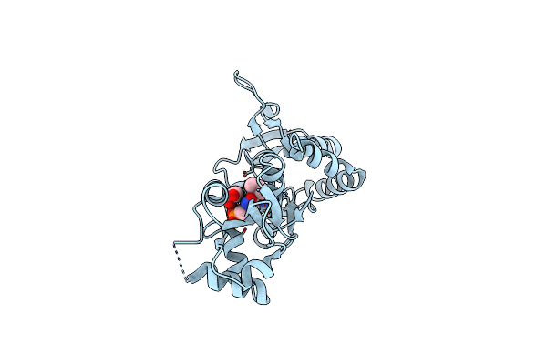
Structure Of The Ampa Receptor Glua2O Ligand-Binding Domain (S1S2J) In Complex With The Compound (S)-1-[2'-Amino-2'-Carboxyethyl]-5,7-Dihydrothieno[3,4-D]Pyrimidin- 2,4(1H,3H)-Dione At Resolution 1.60A
Organism: Rattus norvegicus
Method: X-RAY DIFFRACTION Resolution:1.61 Å Release Date: 2020-06-03 Classification: MEMBRANE PROTEIN Ligands: SO4, GOL, CGW, CL |
 |
Structure Of The Ampa Receptor Glua2O Ligand-Binding Domain (S1S2J) In Complex With The Compound ( S) - 1- [2'-Amino-2'-Carboxyethyl]-5 ,7- Dihydropyrrolo[3,4-D]Pyrimidin-2,4(1H,3H)-Dione At Resolution 1.20A
Organism: Rattus norvegicus
Method: X-RAY DIFFRACTION Resolution:1.20 Å Release Date: 2020-06-03 Classification: MEMBRANE PROTEIN Ligands: GOL, SO4, PVQ, NH4, CL |
 |
Structure Of The Ampa Receptor Glua2O Ligand-Binding Domain (S1S2J) In Complex With The Compound ( S) - 1- [2'-Amino-2'-Carboxyethyl]-6-Methyl-5 ,7- Dihydropyrrolo[3,4-D]Pyrimidin-2,4(1H,3H)-Dione At Resolution 1.00A
Organism: Rattus norvegicus
Method: X-RAY DIFFRACTION Resolution:1.00 Å Release Date: 2020-06-03 Classification: MEMBRANE PROTEIN Ligands: SO4, GOL, LI, CG8, CL |
 |
Structure Of The Ampa Receptor Glua2O Ligand-Binding Domain (S1S2J) In Complex With The Compound (S)-1-(2'-Amino-2'-Carboxyethyl)-5,7-Dihydrofuro[3,4-D]- Pyrimidine-2,4(1H,3H)-Dione At Resolution 1.15A
Organism: Rattus norvegicus
Method: X-RAY DIFFRACTION Resolution:1.15 Å Release Date: 2020-06-03 Classification: MEMBRANE PROTEIN Ligands: GOL, SO4, LI, PVK, CL |
 |
Structure Of The Ampa Receptor Glua2O Ligand-Binding Domain (S1S2J) In Complex With The Compound (S)-1-(2'-Amino-2'-Carboxyethyl)Furo[3,4-D]Pyrimidin-2,4-Dione At Resolution 1.47A
Organism: Rattus norvegicus
Method: X-RAY DIFFRACTION Resolution:1.47 Å Release Date: 2020-06-03 Classification: MEMBRANE PROTEIN Ligands: SO4, GOL, LI, CL, OUB |
 |
Crystal Structure Of The Ligand-Binding Core Of Iglur5 In Complex With The Antagonist (S)-Atpo At 1.85 A Resolution
Organism: Rattus norvegicus
Method: X-RAY DIFFRACTION Resolution:1.85 Å Release Date: 2007-07-03 Classification: MEMBRANE PROTEIN Ligands: AT1, GOL |
 |
Crystal Structure Of The Ligand-Binding Core Of Iglur5 In Complex With The Partial Agonist Domoic Acid At 2.5 A Resolution
Organism: Rattus norvegicus
Method: X-RAY DIFFRACTION Resolution:2.50 Å Release Date: 2007-07-03 Classification: MEMBRANE PROTEIN Ligands: DOQ |
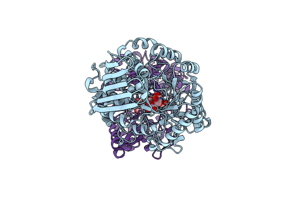 |
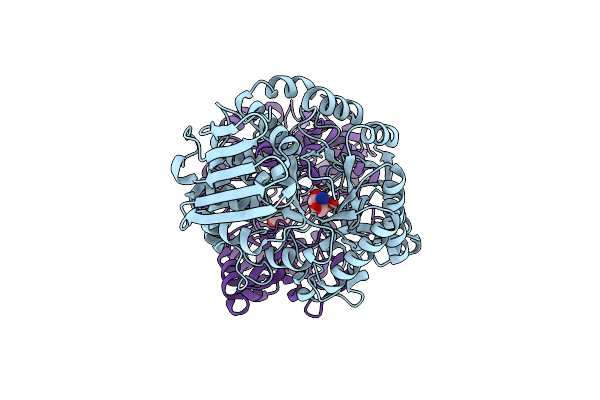
E232Q Mutant Of Sucrose Phosphorylase From Bifidobacterium Adolescentis In Complex With Sucrose
Organism: Bifidobacterium adolescentis
Method: X-RAY DIFFRACTION Resolution:2.10 Å Release Date: 2006-09-26 Classification: TRANSFERASE |
 |
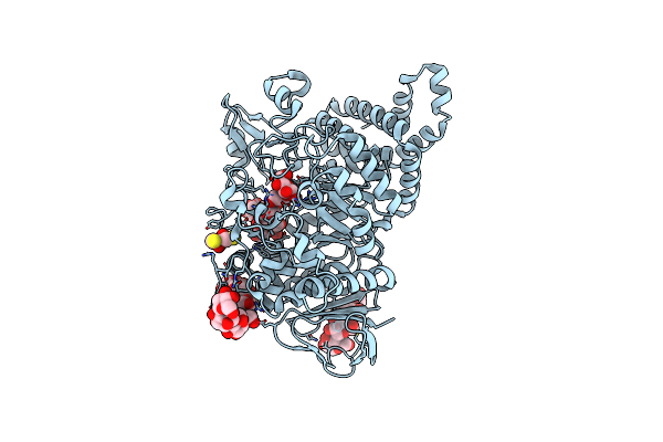
Sucrose Phosphorylase From Bifidobacterium Adolescentis Reacted With Sucrose
Organism: Bifidobacterium adolescentis
Method: X-RAY DIFFRACTION Resolution:2.00 Å Release Date: 2006-09-26 Classification: TRANSFERASE Ligands: BGC |
 |
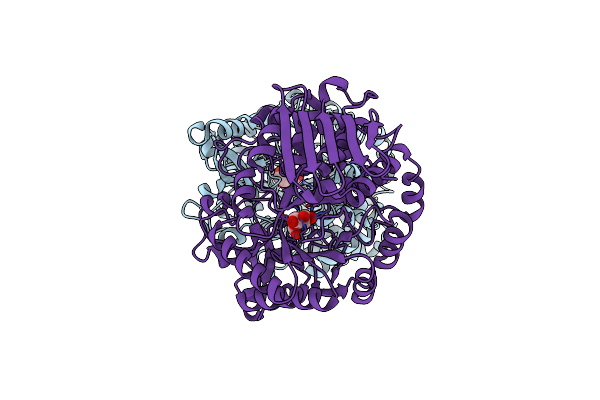
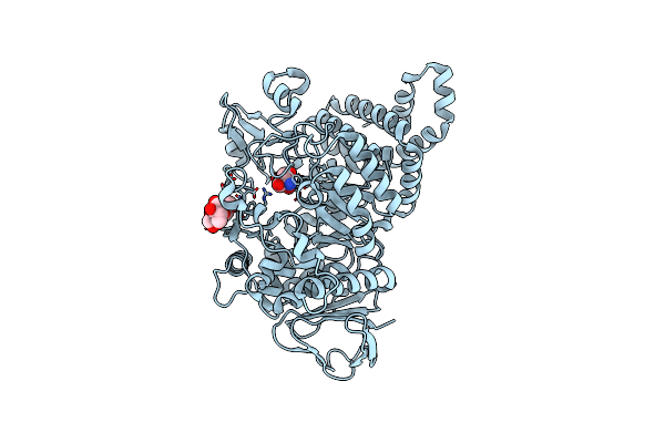
Amylosucrase Mutant E328Q In A Ternary Complex With Sucrose And Maltoheptaose
Organism: Neisseria polysaccharea
Method: X-RAY DIFFRACTION Resolution:2.16 Å Release Date: 2006-05-02 Classification: TRANSFERASE |
 |
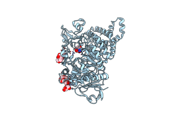
Organism: Bifidobacterium adolescentis
Method: X-RAY DIFFRACTION Resolution:1.77 Å Release Date: 2004-02-10 Classification: TRANSFERASE Ligands: TRS |
 |
Organism: Neisseria polysaccharea
Method: X-RAY DIFFRACTION Resolution:2.00 Å Release Date: 2002-12-18 Classification: TRANSFERASE Ligands: TRS |
 |
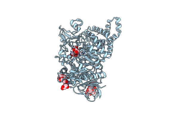
Amylosucrase Mutant E328Q Co-Crystallized With Maltoheptaose Then Soaked With Maltoheptaose.
Organism: Neisseria polysaccharea
Method: X-RAY DIFFRACTION Resolution:2.01 Å Release Date: 2002-12-18 Classification: TRANSFERASE Ligands: DTT |
 |
Organism: Neisseria polysaccharea
Method: X-RAY DIFFRACTION Resolution:2.10 Å Release Date: 2002-12-18 Classification: TRANSFERASE Ligands: TRS |
 |
Organism: Neisseria polysaccharea
Method: X-RAY DIFFRACTION Resolution:2.10 Å Release Date: 2002-12-18 Classification: TRANSFERASE Ligands: TRS |
 |
Organism: Neisseria polysaccharea
Method: X-RAY DIFFRACTION Resolution:2.00 Å Release Date: 2002-12-18 Classification: TRANSFERASE Ligands: TRS |