
Search Count: 16
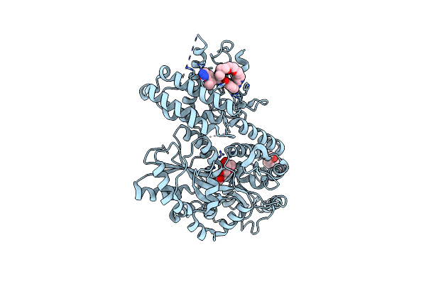 |
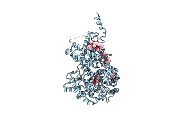
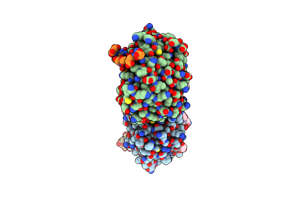
Crystal Structure Of A Cgrp Receptor Ectodomain Heterodimer Bound To Macrocyclic Inhibitor Htl0029881
Organism: Escherichia coli k-12, Homo sapiens
Method: X-RAY DIFFRACTION Resolution:2.75 Å Release Date: 2022-12-07 Classification: MEMBRANE PROTEIN Ligands: OKU, PG4 |
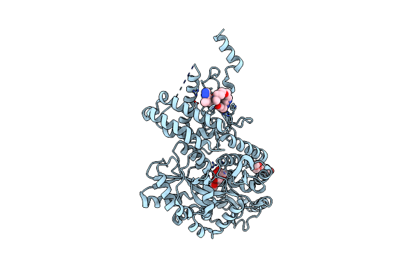 |
Crystal Structure Of A Cgrp Receptor Ectodomain Heterodimer Bound To Macrocyclic Inhibitor Htl0029882
Organism: Escherichia coli k-12, Homo sapiens
Method: X-RAY DIFFRACTION Resolution:1.90 Å Release Date: 2022-12-07 Classification: MEMBRANE PROTEIN Ligands: OP9, PG4 |
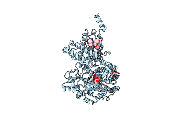 |
Crystal Structure Of A Cgrp Receptor Ectodomain Heterodimer Bound To Macrocyclic Inhibitor Htl0031448
Organism: Escherichia coli k-12, Homo sapiens
Method: X-RAY DIFFRACTION Resolution:1.65 Å Release Date: 2022-12-07 Classification: MEMBRANE PROTEIN Ligands: PG4, OL0, ACT |
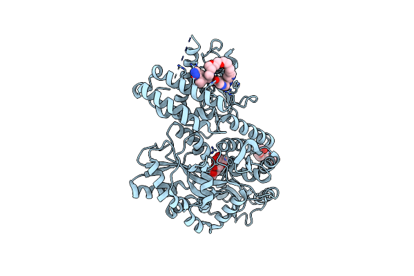 |
Crystal Structure Of A Cgrp Receptor Ectodomain Heterodimer Bound To Macrocyclic Inhibitor Htl0028125
Organism: Escherichia coli (strain k12), Homo sapiens
Method: X-RAY DIFFRACTION Resolution:1.85 Å Release Date: 2022-06-15 Classification: MEMBRANE PROTEIN Ligands: 7IR, PG4 |
 |
Crystal Structure Of A Cgrp Receptor Ectodomain Heterodimer Bound To Macrocyclic Inhibitor Compound 13
Organism: Escherichia coli (strain k12), Homo sapiens
Method: X-RAY DIFFRACTION Resolution:2.30 Å Release Date: 2022-06-15 Classification: MEMBRANE PROTEIN Ligands: PG4, 7IU |
 |
Crystal Structure Of A Cgrp Receptor Ectodomain Heterodimer With Bound High Affinity Inhibitor
Organism: Escherichia coli (strain k12), Homo sapiens
Method: X-RAY DIFFRACTION Resolution:1.60 Å Release Date: 2020-07-15 Classification: MEMBRANE PROTEIN Ligands: QLQ, PG4 |
 |
Crystal Structure Of A Cgrp Receptor Ectodomain Heterodimer With Bound High Affinity Inhibitor
Organism: Escherichia coli (strain k12), Homo sapiens
Method: X-RAY DIFFRACTION Resolution:1.73 Å Release Date: 2020-07-15 Classification: MEMBRANE PROTEIN Ligands: PG4, 3N6 |
 |
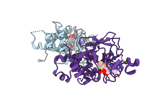
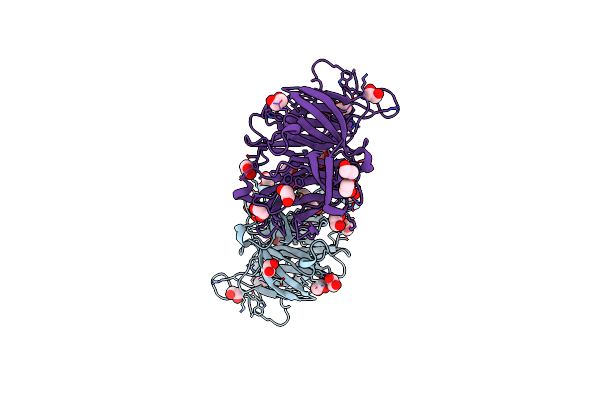
Crystal Structure Of The Human Glucagon Receptor (Gcgr) In Complex With The Antagonist Mk-0893
Organism: Homo sapiens, Enterobacteria phage t4
Method: X-RAY DIFFRACTION Resolution:2.50 Å Release Date: 2016-04-20 Classification: SIGNALING PROTEIN Ligands: 5MV, OLA, PE5, TLA |
 |
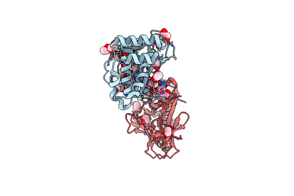
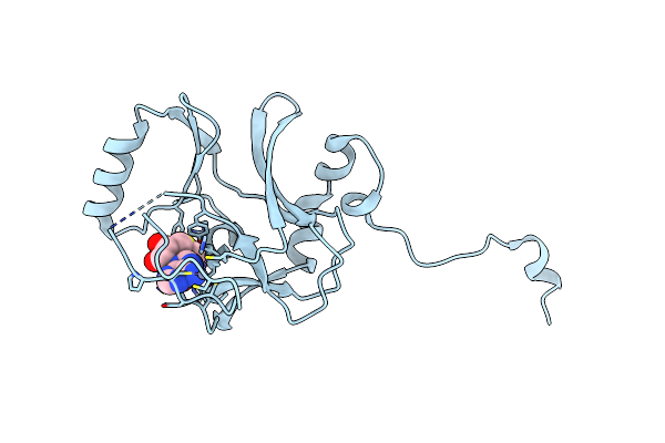
A Novel Route To Product Specificity In The Suv4-20 Family Of Histone H4K20 Methyltransferases
Organism: Mus musculus
Method: X-RAY DIFFRACTION Resolution:2.17 Å Release Date: 2013-10-02 Classification: TRANSFERASE Ligands: ZN, SAM, GOL |
 |
Organism: Mus musculus
Method: X-RAY DIFFRACTION Release Date: 2013-05-22 Classification: TRANSFERASE Ligands: SAH, ZN, EDO |
 |
Organism: Mus musculus
Method: X-RAY DIFFRACTION Resolution:2.40 Å Release Date: 2010-08-04 Classification: TRANSCRIPTION Ligands: GOL |
 |
Organism: Mus musculus
Method: X-RAY DIFFRACTION Resolution:2.70 Å Release Date: 2010-08-04 Classification: TRANSCRIPTION Ligands: GOL |
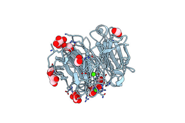 |
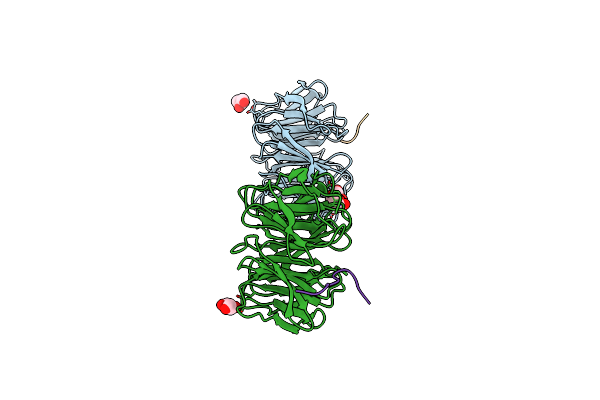
Structure Of The Pqq-Bound Form Of Aldose Sugar Dehydrogenase (Adh) From Streptomyces Coelicolor
Organism: Streptomyces coelicolor
Method: X-RAY DIFFRACTION Resolution:1.60 Å Release Date: 2009-06-02 Classification: OXIDOREDUCTASE Ligands: ARA, CA, PQQ, EDO |
 |
Binary Complex Of The Mixed Lineage Leukaemia (Mll1) Set Domain With The Cofactor Product S-Adenosylhomocysteine.
Organism: Homo sapiens
Method: X-RAY DIFFRACTION Release Date: 2009-02-10 Classification: TRANSFERASE Ligands: ZN, SAH |
 |
Ternary Complex Of The Mixed Lineage Leukaemia (Mll1) Set Domain With The Cofactor Product S-Adenosylhomocysteine And Histone Peptide.
Organism: Homo sapiens
Method: X-RAY DIFFRACTION Resolution:2.20 Å Release Date: 2009-02-10 Classification: TRANSFERASE Ligands: ZN, SAH, GOL |
 |
Crystal Structure Of The Soluble Aldose Sugar Dehydrogenase (Asd) From Escherichia Coli In The Apo-Form
Organism: Escherichia coli k12
Method: X-RAY DIFFRACTION Resolution:1.50 Å Release Date: 2006-08-08 Classification: SUGAR BINDING PROTEIN Ligands: CA, PO4, EDO |