
Search Count: 8
 |
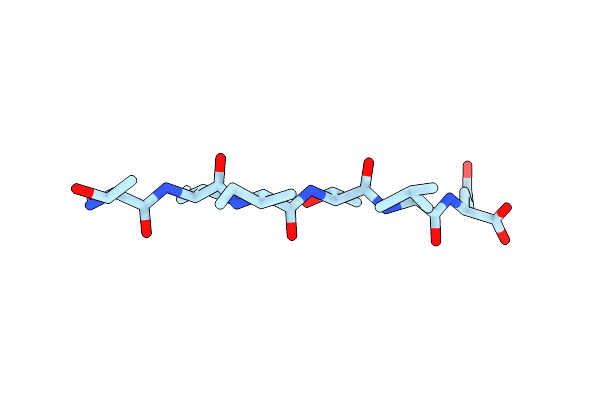
Structure Of The Amyloid-Forming Segment Ltiitle From P53 (Residues 252-258)
Organism: Homo sapiens
Method: X-RAY DIFFRACTION Resolution:1.70 Å Release Date: 2016-01-13 Classification: PROTEIN FIBRIL |
 |
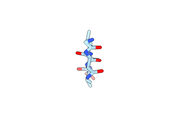
Structure Of The Amyloid-Forming Segment Tiitle From P53 (Residues 253-258)
Organism: Homo sapiens
Method: X-RAY DIFFRACTION Resolution:1.58 Å Release Date: 2016-01-13 Classification: PROTEIN FIBRIL Ligands: ZN |
 |
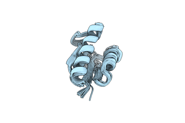
Structure Of The Amyloid Forming Peptide Gnlvs (Residues 26-30) From The Eosinophil Major Basic Protein (Embp)
Organism: Homo sapiens
Method: X-RAY DIFFRACTION Resolution:1.45 Å Release Date: 2015-03-18 Classification: PROTEIN FIBRIL |
 |
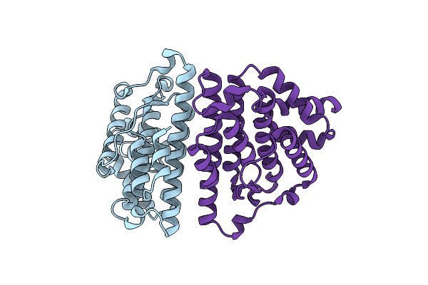
Organism: Podospora anserina
Method: SOLID-STATE NMR Release Date: 2011-06-01 Classification: PROTEIN FIBRIL Ligands: CGO |
 |
Organism: Podospora anserina
Method: X-RAY DIFFRACTION Resolution:2.62 Å Release Date: 2010-07-28 Classification: PRION-BINDING PROTEIN |
 |
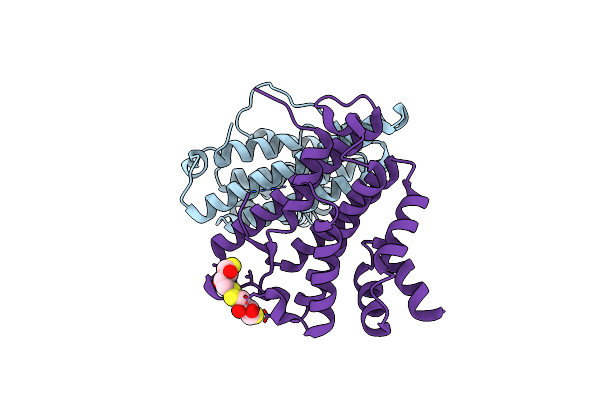
Organism: Podospora anserina
Method: X-RAY DIFFRACTION Resolution:2.30 Å Release Date: 2010-07-28 Classification: PRION-BINDING PROTEIN Ligands: CL |
 |
Organism: Podospora anserina
Method: X-RAY DIFFRACTION Resolution:2.00 Å Release Date: 2010-07-28 Classification: PRION-BINDING PROTEIN Ligands: DTT, DTU |
 |