
Search Count: 546
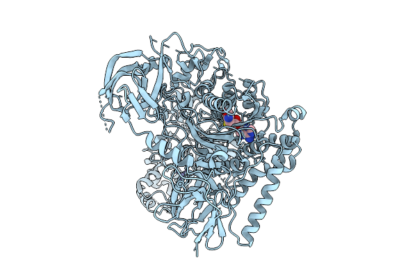 |
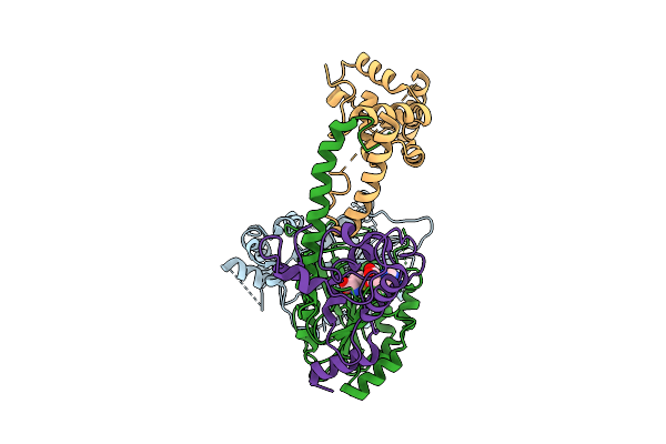
Organism: Arabidopsis thaliana
Method: ELECTRON MICROSCOPY Release Date: 2025-10-22 Classification: TRANSFERASE Ligands: SAH, ZN |
 |
Organism: Arabidopsis thaliana
Method: ELECTRON MICROSCOPY Release Date: 2025-10-22 Classification: TRANSFERASE Ligands: ZN, SAH |
 |
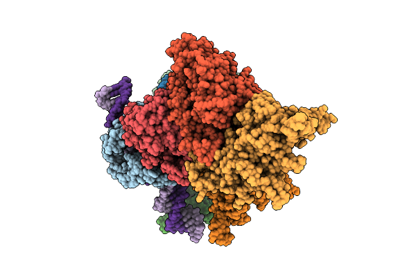
Organism: Arabidopsis thaliana, Synthetic construct
Method: ELECTRON MICROSCOPY Release Date: 2025-10-22 Classification: TRANSFERASE/DNA Ligands: ZN |
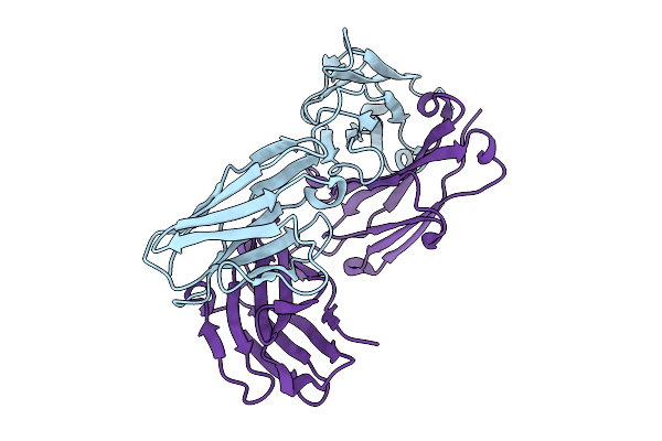 |
Organism: Mus musculus
Method: X-RAY DIFFRACTION Release Date: 2025-08-27 Classification: IMMUNE SYSTEM |
 |
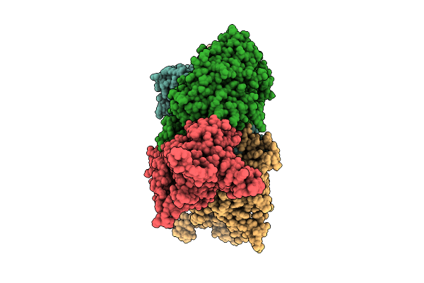
Organism: Homo sapiens, Oryctolagus cuniculus
Method: ELECTRON MICROSCOPY Release Date: 2025-08-13 Classification: CYTOSOLIC PROTEIN/CONTRACTILE PROTEIN |
 |
Organism: Homo sapiens, Oryctolagus cuniculus
Method: ELECTRON MICROSCOPY Release Date: 2025-08-13 Classification: CYTOSOLIC PROTEIN/CONTRACTILE PROTEIN |
 |
 |
Organism: Salmonella phage pjns002
Method: ELECTRON MICROSCOPY Release Date: 2025-08-13 Classification: VIRUS |
 |
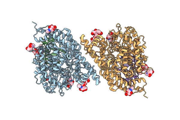
Organism: Tetrahymena thermophila sb210
Method: ELECTRON MICROSCOPY Release Date: 2025-08-13 Classification: TRANSFERASE Ligands: SAH |
 |
Structure Of Hku5 Spike C-Terminal Domain In Complex With Ace2 From Pipistrellus Abramus
Organism: Pipistrellus abramus, Pipistrellus bat coronavirus hku5
Method: ELECTRON MICROSCOPY Release Date: 2025-07-30 Classification: VIRAL PROTEIN Ligands: NAG |
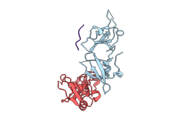 |
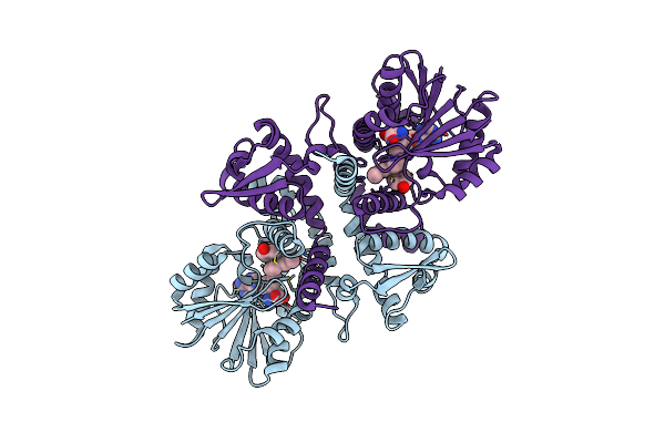
Crystal Structure Of Mouse Bahcc1 Ttd Domain In Complex With H4K20Me1 Peptide
Organism: Mus musculus, Homo sapiens
Method: X-RAY DIFFRACTION Release Date: 2025-07-16 Classification: PROTEIN BINDING |
 |
Organism: Homo sapiens
Method: ELECTRON MICROSCOPY Release Date: 2025-07-09 Classification: STRUCTURAL PROTEIN Ligands: ZN |
 |
Organism: Homo sapiens
Method: ELECTRON MICROSCOPY Release Date: 2025-07-09 Classification: STRUCTURAL PROTEIN Ligands: ZN |
 |
Organism: Bovine adenovirus 3
Method: ELECTRON MICROSCOPY Release Date: 2025-06-04 Classification: VIRAL PROTEIN |
 |
The Crystal Structure Of An Atypical N-Methyltransferasea Paomt9 In P. Amurense
Organism: Phellodendron amurense
Method: X-RAY DIFFRACTION Release Date: 2025-05-28 Classification: TRANSFERASE Ligands: SAM, A1D8J |
 |
Crystal Structure Of Zmet2 In Complex With Unmethylated Ctg Dna And A Histone H3Kc9Me2 Peptide
Organism: Zea mays, Synthetic construct
Method: X-RAY DIFFRACTION Resolution:2.71 Å Release Date: 2025-05-21 Classification: TRANSFERASE Ligands: SAH |
 |
Crystal Structure Of Enl Yeats In Complex With Histone H3 Methacrylated At K18
Organism: Homo sapiens
Method: X-RAY DIFFRACTION Release Date: 2025-04-09 Classification: TRANSCRIPTION Ligands: MES |
 |
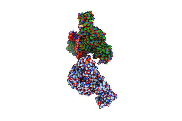
Codanin-1 Sequesters Asf1 By Using A Histone H3 Mimic Helix To Regulate Histone Supply
Organism: Homo sapiens
Method: ELECTRON MICROSCOPY Release Date: 2025-03-19 Classification: REPLICATION |
 |
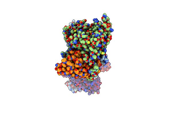
Organism: Homo sapiens
Method: ELECTRON MICROSCOPY Release Date: 2025-03-12 Classification: GENE REGULATION Ligands: ZN |
 |
Organism: Escherichia phage t1
Method: ELECTRON MICROSCOPY Release Date: 2025-03-12 Classification: VIRAL PROTEIN |