
Search Count: 43
 |
 |
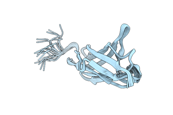
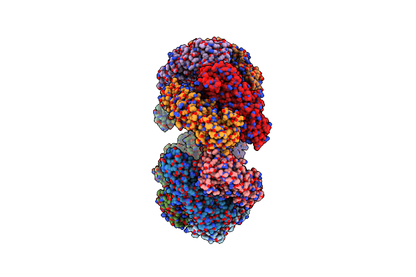
Organism: Mus musculus, Rotavirus a
Method: ELECTRON MICROSCOPY Release Date: 2025-02-12 Classification: VIRUS |
 |
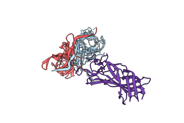
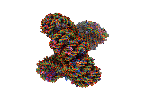
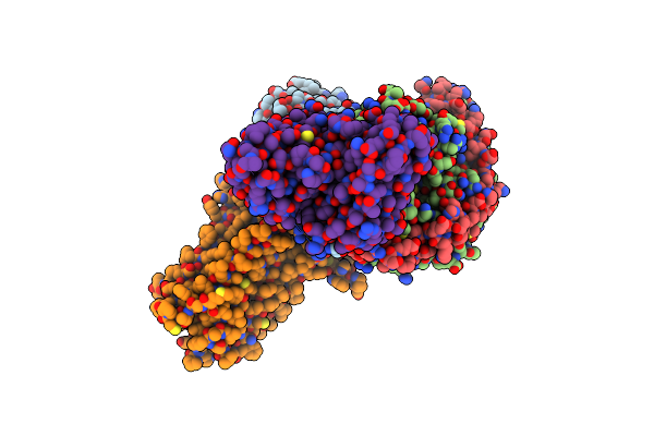
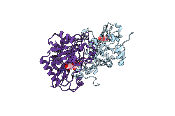
Cryo-Em Structure Of Simian Rotavirus Sa11 Vp4 In Complex With Nab 7H13-I54G Mutant (Left Side)
Organism: Mus musculus, Rotavirus a
Method: ELECTRON MICROSCOPY Release Date: 2025-02-12 Classification: VIRUS |
 |
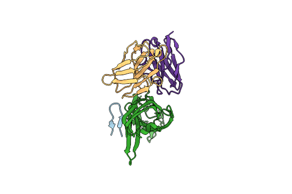
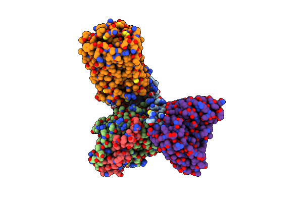
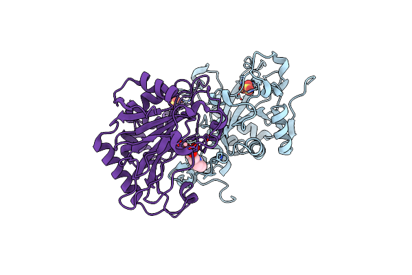
Cryo-Em Structure Of Simian Rotavirus Sa11 Vp4 In Complex With Nab 7H13-I54G Mutant (Right Side)
Organism: Mus musculus, Rotavirus a
Method: ELECTRON MICROSCOPY Release Date: 2025-02-12 Classification: VIRUS |
 |
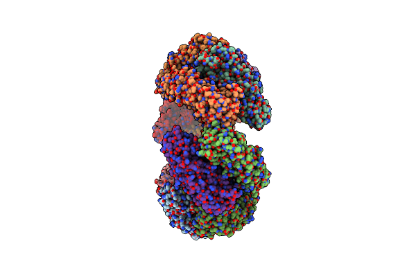
Structural Basis For The Linker Histone H5-Nucleosome Binding And Chromatin Compaction
Organism: Synthetic construct, Xenopus laevis, Gallus gallus
Method: ELECTRON MICROSCOPY Release Date: 2024-09-11 Classification: GENE REGULATION |
 |
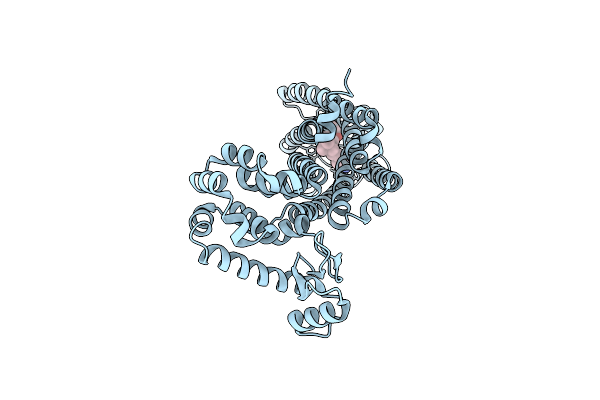
Organism: Homo sapiens
Method: ELECTRON MICROSCOPY Release Date: 2020-04-15 Classification: TRANSPORT PROTEIN |
 |
Organism: Homo sapiens
Method: ELECTRON MICROSCOPY Release Date: 2020-04-15 Classification: TRANSPORT PROTEIN |
 |
Organism: Homo sapiens
Method: ELECTRON MICROSCOPY Release Date: 2020-04-15 Classification: TRANSPORT PROTEIN |
 |
Organism: Homo sapiens, Enterobacteria phage rb59
Method: X-RAY DIFFRACTION Resolution:3.20 Å Release Date: 2020-02-12 Classification: MEMBRANE PROTEIN Ligands: E3R |
 |
Organism: Homo sapiens
Method: ELECTRON MICROSCOPY Release Date: 2020-02-12 Classification: MEMBRANE PROTEIN Ligands: E3R |
 |
Organism: Homo sapiens
Method: ELECTRON MICROSCOPY Release Date: 2020-02-12 Classification: MEMBRANE PROTEIN Ligands: 8D0 |
 |
Organism: Chenopodium quinoa
Method: SOLUTION NMR Release Date: 2019-05-15 Classification: PLANT PROTEIN |
 |
 |
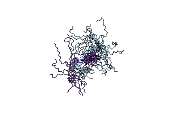
Organism: Homo sapiens
Method: SOLUTION NMR Release Date: 2018-05-02 Classification: SIGNALING PROTEIN |
 |
Organism: Homo sapiens
Method: SOLUTION NMR Release Date: 2018-05-02 Classification: SIGNALING PROTEIN |
 |
 |
Organism: Neosartorya fumigata
Method: X-RAY DIFFRACTION Resolution:2.54 Å Release Date: 2015-11-04 Classification: OXIDOREDUCTASE Ligands: AKG, FE2, CO |
 |
Organism: Neosartorya fumigata
Method: X-RAY DIFFRACTION Resolution:1.95 Å Release Date: 2015-11-04 Classification: OXIDOREDUCTASE Ligands: FE2, SO4, CO, MES |
 |
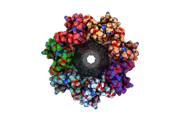
Crystal Structure Of Dodecameric Iron-Containing Heptosyltransferase Tibc In Complex With Adp-D-Beta-D-Heptose At 3.9 Angstrom Resolution
Organism: Escherichia coli dec13e
Method: X-RAY DIFFRACTION Resolution:3.88 Å Release Date: 2014-11-05 Classification: TRANSFERASE Ligands: FE, AQH, EDO |
 |
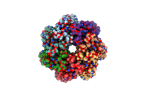
Crystal Structure Of Bacterial Iron-Containing Dodecameric Glycosyltransferase Tibc From Enterotoxigenic E.Coli H10407
Organism: Escherichia coli etec h10407
Method: X-RAY DIFFRACTION Resolution:2.88 Å Release Date: 2014-10-29 Classification: TRANSFERASE Ligands: FE, EDO |