Planned Maintenance: Some services may turn out to be unavailable from 15th January, 2026 to 16th January, 2026. We apologize for the inconvenience!

Planned Maintenance: Some services may turn out to be unavailable from 15th January, 2026 to 16th January, 2026. We apologize for the inconvenience!
 |
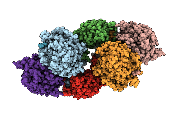
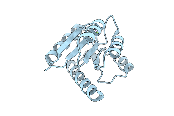
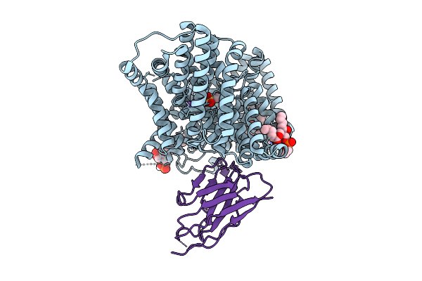
Human Ectonucleotide Pyrophosphatase/Phosphodiesterase Family Member 3 (Enpp3) Inhibitor Complex
Organism: Homo sapiens
Method: X-RAY DIFFRACTION Release Date: 2026-01-07 Classification: HYDROLASE Ligands: A1BYU, ZN, CA, NAG, CL |
 |
[Fefe]-Hydrogenase From D. Desulfuricans With Synthetic Active Site Containing Only One Cyanide Ligand.
Organism: Desulfovibrio desulfuricans
Method: X-RAY DIFFRACTION Release Date: 2025-12-24 Classification: OXIDOREDUCTASE Ligands: SF4, A1IW2, CL, LI |
 |
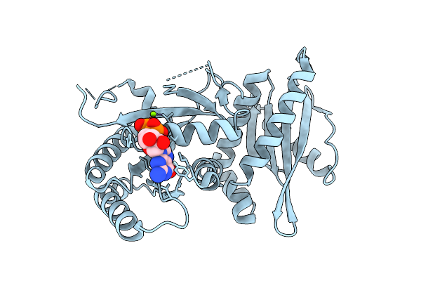
Organism: Homo sapiens
Method: X-RAY DIFFRACTION Release Date: 2025-12-24 Classification: HYDROLASE/INHIBITOR Ligands: A1C2U, EDO |
 |
Organism: Avian orthoreovirus
Method: ELECTRON MICROSCOPY Release Date: 2025-12-03 Classification: VIRAL PROTEIN |
 |
Crystal Structure Of Monomeric Rag-Like Small Gtpase From Asgard Lokiarchaeota (Lokiragm) In Complex With Gdp
Organism: Candidatus prometheoarchaeum syntrophicum
Method: X-RAY DIFFRACTION Release Date: 2025-11-26 Classification: HYDROLASE Ligands: GDP, MG |
 |
Structure Of The Acinetobacter Baumannii Response Regulator Pmra Receiver Domain D10N Mutation
Organism: Acinetobacter baumannii
Method: X-RAY DIFFRACTION Release Date: 2025-11-26 Classification: TRANSCRIPTION Ligands: MG |
 |
Structure Of The Acinetobacter Baumannii Response Regulator Pmra Receiver Domain M12I Mutation
Organism: Acinetobacter baumannii
Method: X-RAY DIFFRACTION Release Date: 2025-11-26 Classification: TRANSCRIPTION Ligands: MG |
 |
Structure Of The Acinetobacter Baumannii Response Regulator Pmra Receiver Domain I13M Mutation
Organism: Acinetobacter baumannii
Method: X-RAY DIFFRACTION Release Date: 2025-11-26 Classification: TRANSCRIPTION Ligands: MG |
 |
Structure Of The Acinetobacter Baumannii Response Regulator Pmra Receiver Domain I13M Mutation In Active Dimer State
Organism: Acinetobacter baumannii
Method: X-RAY DIFFRACTION Release Date: 2025-11-26 Classification: TRANSCRIPTION |
 |
Structure Of The Acinetobacter Baumannii Response Regulator Pmra Receiver Domain G54E Mutation
Organism: Acinetobacter baumannii
Method: X-RAY DIFFRACTION Release Date: 2025-11-26 Classification: TRANSCRIPTION Ligands: MG |
 |
Structure Of The Acinetobacter Baumannii Response Regulator Pmra Receiver Domain S119T Mutation
Organism: Acinetobacter baumannii
Method: X-RAY DIFFRACTION Release Date: 2025-11-26 Classification: TRANSCRIPTION |
 |
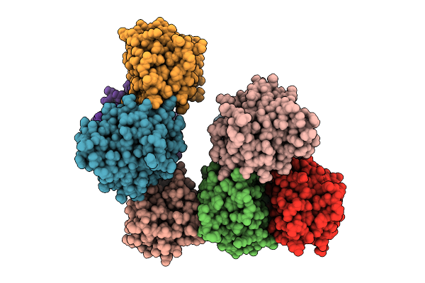
Cryo-Em Structure Of The Isethionate Trap Transporter Iseqm From Oleidesulfovibrio Alaskensis With Bound Isethionate
Organism: Oleidesulfovibrio alaskensis g20, Helicobacter pylori
Method: ELECTRON MICROSCOPY Release Date: 2025-11-12 Classification: TRANSPORT PROTEIN |
 |
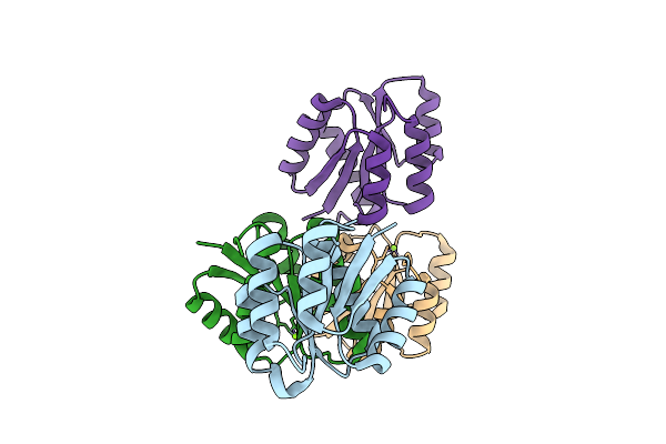
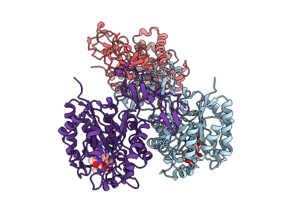
Organism: Homo sapiens
Method: ELECTRON MICROSCOPY Release Date: 2025-11-12 Classification: OXIDOREDUCTASE Ligands: NDP, HEM |
 |
Organism: Agrobacterium radiobacter
Method: ELECTRON MICROSCOPY Release Date: 2025-11-12 Classification: OXIDOREDUCTASE Ligands: HEM, NDP |
 |
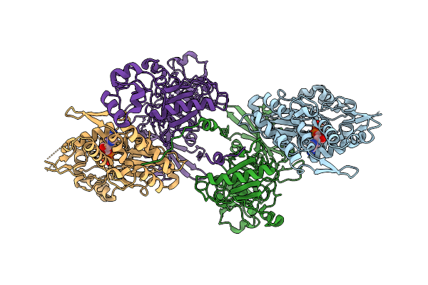
Catalase Cryoem Structure From Micrococcus Luteus At 1.9 Angstrom Resolution.
Organism: Micrococcus luteus
Method: ELECTRON MICROSCOPY Release Date: 2025-11-12 Classification: OXIDOREDUCTASE Ligands: HEM, NDP |
 |
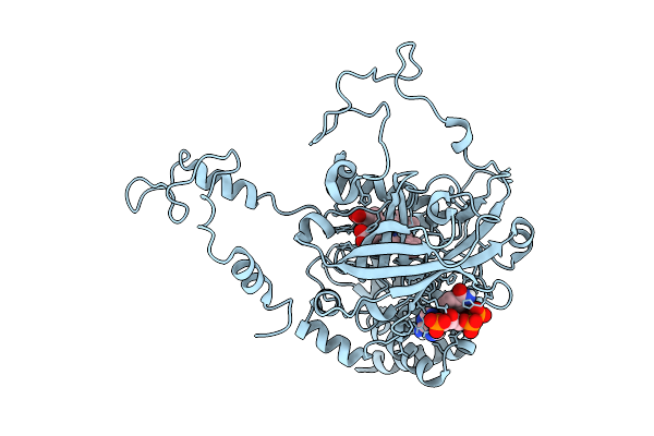
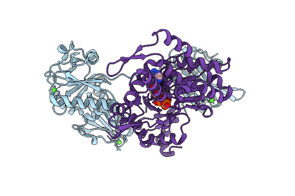
Crystal Structure Of Kirsten Rat Sarcoma G12C Complexed With Gmppnp And Covalently Bound To An Adduct Of {(2S)-4-[7-(8-Chloronaphthalen-1-Yl)-2-{[(2S)-1-Methylpyrrolidin-2-Yl]Methoxy}-5,6,7,8-Tetrahydropyrido[3,4-D]Pyrimidin-4-Yl]-1-[(2Z)-2-Fluoro-3-(Pyridin-2-Yl)Prop-2-Enoyl]Piperazin-2-Yl}Acetonitrile
Organism: Homo sapiens
Method: X-RAY DIFFRACTION Release Date: 2025-11-05 Classification: HYDROLASE/HYDROLASE INHIBITOR Ligands: GNP, A1B7P, MG |
 |
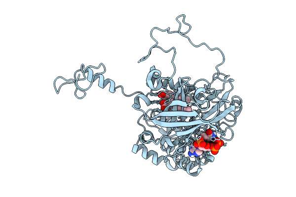
Crystal Structure Of Kirsten Rat Sarcoma G12C Complexed With Gdp And Covalently Bound To An Adduct Of (2S)-1-{4-[(7P)-7-(8-Ethynyl-7-Fluoro-3-Hydroxynaphthalen-1-Yl)-8-Fluoro-2-{[(2R,4R,7As)-2-Fluorotetrahydro-1H-Pyrrolizin-7A(5H)-Yl]Methoxy}Pyrido[4,3-D]Pyrimidin-4-Yl]Piperazin-1-Yl}-2-Fluoro-3-(1,3-Thiazol-2-Yl)Propan-1-One
Organism: Homo sapiens
Method: X-RAY DIFFRACTION Release Date: 2025-11-05 Classification: HYDROLASE/HYDROLASE INHIBITOR Ligands: GDP, A1B7Q, MG |
 |
Organism: Paralvinella sulfincola, Gallus gallus
Method: X-RAY DIFFRACTION Release Date: 2025-10-15 Classification: STRUCTURAL PROTEIN Ligands: CA, ADP |
 |
Organism: Paralvinella sulfincola, Gallus gallus
Method: X-RAY DIFFRACTION Release Date: 2025-10-15 Classification: STRUCTURAL PROTEIN Ligands: CA, ATP |
 |
Chito Oligosaccharide Deacetylase From Vibrio Campbellii (Vhcod) In Complex With Triacetyl-Chitotriose (Glcnac)3
Organism: Vibrio campbellii atcc baa-1116
Method: X-RAY DIFFRACTION Release Date: 2025-10-08 Classification: HYDROLASE Ligands: ZN |