
Search Count: 4,313
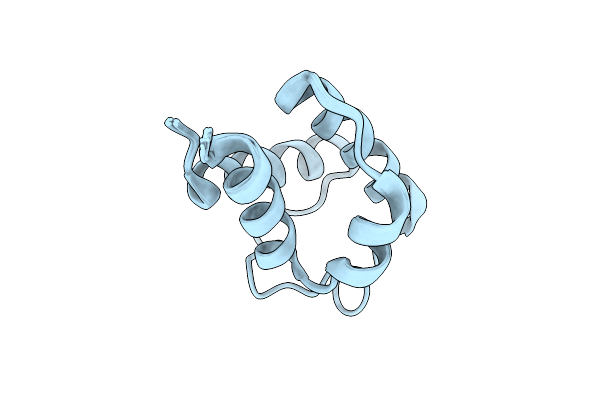 |
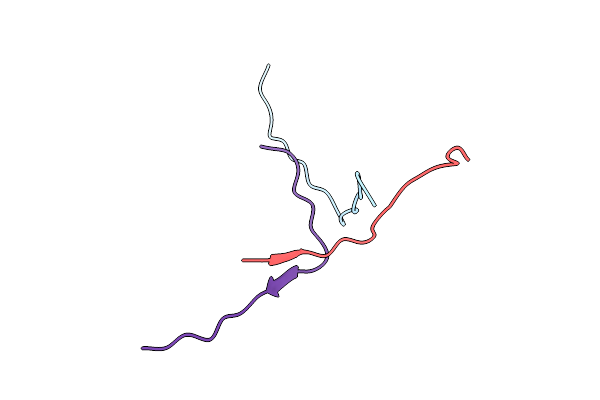
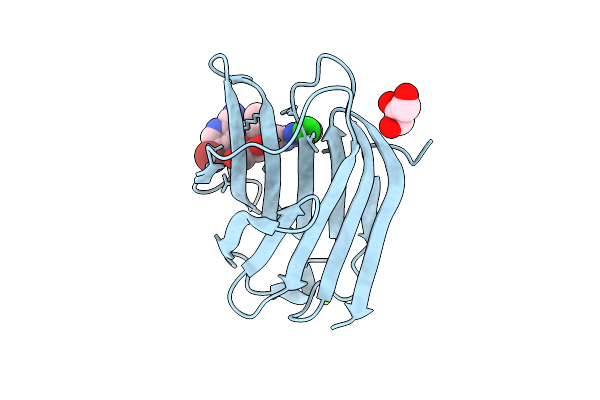
Solution Structure Of Holo Acyl Carrier Protein 2 (Apef) Of Aryl Polyene Biosynthesis From Acinetobacter Baumannii
Organism: Acinetobacter baumannii
Method: SOLUTION NMR Release Date: 2025-11-26 Classification: BIOSYNTHETIC PROTEIN |
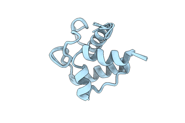 |
Solution Structure Of Holo Acyl Carrier Protein 1 (Apee) Of Aryl Polyene Biosynthesis From Acinetobacter Baumannii
Organism: Acinetobacter baumannii
Method: SOLUTION NMR Release Date: 2025-11-26 Classification: BIOSYNTHETIC PROTEIN |
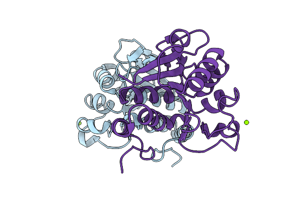 |
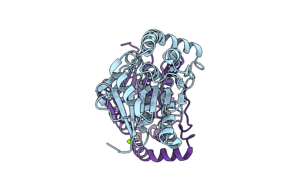
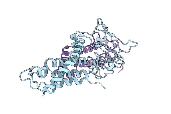
Organism: Escherichia coli
Method: X-RAY DIFFRACTION Release Date: 2025-11-26 Classification: HYDROLASE Ligands: MG, CL |
 |
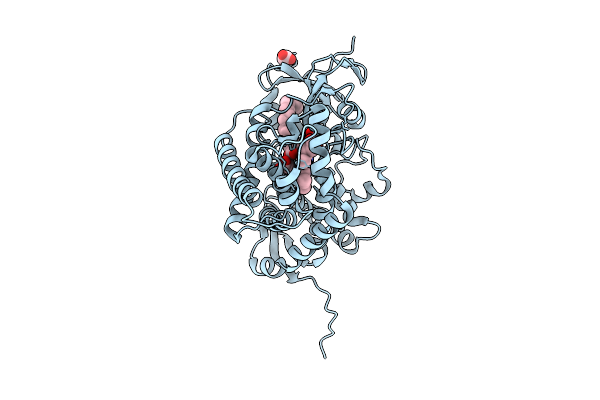
Organism: Homo sapiens
Method: X-RAY DIFFRACTION, NEUTRON DIFFRACTION Release Date: 2025-11-19 Classification: HYDROLASE Ligands: D8U |
 |
Organism: Escherichia coli
Method: X-RAY DIFFRACTION, NEUTRON DIFFRACTION Release Date: 2025-11-19 Classification: HYDROLASE Ligands: D8U |
 |
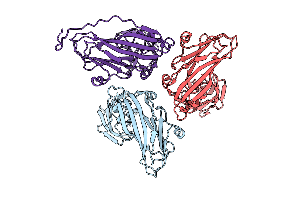
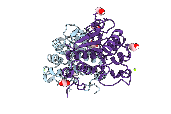
Cytochrome P450 Decarboxylase From Staphylococcus Aureus (Olet_Sa) With Elaidic Acid And Acetate Bound
Organism: Staphylococcus aureus
Method: X-RAY DIFFRACTION Release Date: 2025-11-12 Classification: OXIDOREDUCTASE Ligands: HEM, ELA, ACT |
 |
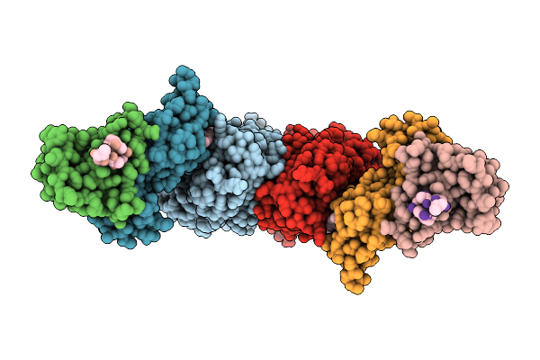
Icosahedral Symmetric Structure Of An Expansion Intermediate Of Turnip Crinkle Virus (Asymmetric Trimer Unit)
Organism: Turnip crinkle virus
Method: ELECTRON MICROSCOPY Release Date: 2025-11-12 Classification: VIRUS |
 |
Symmetry Relaxed Asymmetric Structure Of An Expansion Intermediate Of Turnip Crinkle Virus
Organism: Turnip crinkle virus
Method: ELECTRON MICROSCOPY Release Date: 2025-11-12 Classification: VIRAL PROTEIN |
 |
Organism: Homo sapiens
Method: ELECTRON MICROSCOPY Release Date: 2025-11-05 Classification: TRANSFERASE Ligands: A1BWF |
 |
Organism: Pseudomonas fluorescens
Method: X-RAY DIFFRACTION Release Date: 2025-10-29 Classification: LYASE Ligands: EDO, MES |
 |
Organism: Pseudomonas fluorescens
Method: X-RAY DIFFRACTION Release Date: 2025-10-29 Classification: LYASE Ligands: MG, CL |
 |
Organism: Pseudomonas fluorescens
Method: X-RAY DIFFRACTION Release Date: 2025-10-29 Classification: LYASE Ligands: CL |
 |
Organism: Homo sapiens, Mus musculus
Method: ELECTRON MICROSCOPY Release Date: 2025-10-22 Classification: TRANSFERASE Ligands: ZN |
 |
Organism: Escherichia coli
Method: X-RAY DIFFRACTION Release Date: 2025-10-22 Classification: HYDROLASE Ligands: EDO, CL, MG |
 |
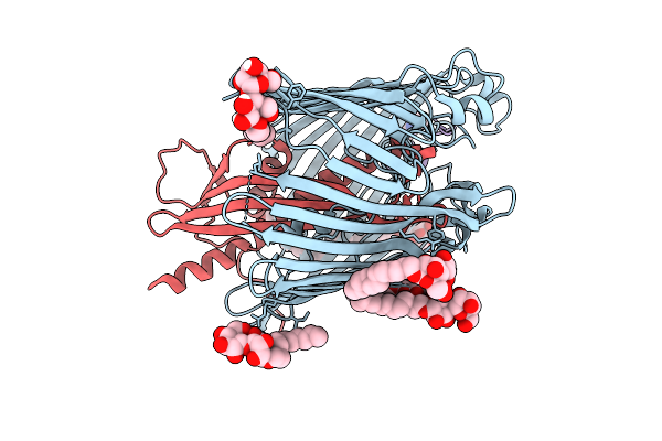
Organism: Homo sapiens
Method: X-RAY DIFFRACTION Release Date: 2025-10-15 Classification: SUGAR BINDING PROTEIN Ligands: YNO, GOL, MG, CL |
 |
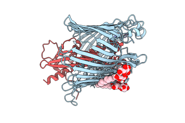
Cryo-Em Structure Of Shigella Flexneri Lptde Bound By A Bicyclic Peptide Molecule (Compound 1)
Organism: Shigella flexneri, Synthetic construct
Method: ELECTRON MICROSCOPY Release Date: 2025-10-15 Classification: MEMBRANE PROTEIN Ligands: LMT, A1I1D |
 |
Cryo-Em Structure Of Shigella Flexneri Lptde Bound By A Bicyclic Peptide Molecule (Compound 2)
Organism: Shigella flexneri, Synthetic construct
Method: ELECTRON MICROSCOPY Release Date: 2025-10-15 Classification: MEMBRANE PROTEIN Ligands: LMT, KZ0 |
 |
Cryo-Em Structure Of Shigella Flexneri Lptde Bound By A Bicyclic Peptide Molecule (Compound 3)
Organism: Shigella flexneri, Synthetic construct
Method: ELECTRON MICROSCOPY Release Date: 2025-10-15 Classification: MEMBRANE PROTEIN Ligands: LMT, KZ0 |
 |
Cryo-Em Structure Of Shigella Flexneri Lptde Bound By A Bicyclic Peptide Molecule (Compound 4)
Organism: Shigella flexneri, Synthetic construct
Method: ELECTRON MICROSCOPY Release Date: 2025-10-15 Classification: MEMBRANE PROTEIN Ligands: LMT, A1I1E |
 |
Cryo-Em Structure Of Shigella Flexneri Lptde Bound By A Bicyclic Peptide Molecule (Compound 5)
Organism: Shigella flexneri, Synthetic construct
Method: ELECTRON MICROSCOPY Release Date: 2025-10-15 Classification: MEMBRANE PROTEIN Ligands: LMT, R06 |