
Search Count: 48
 |
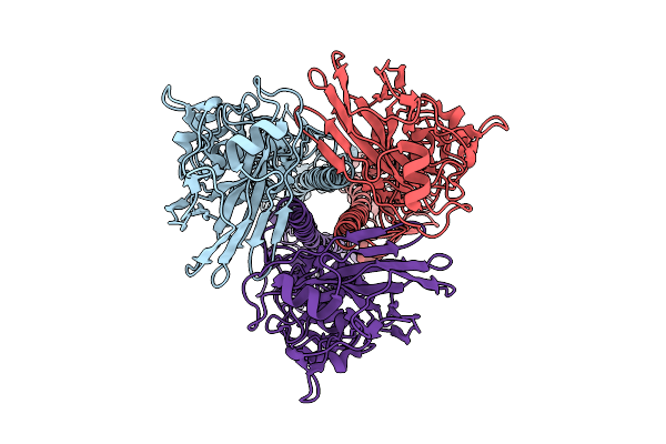
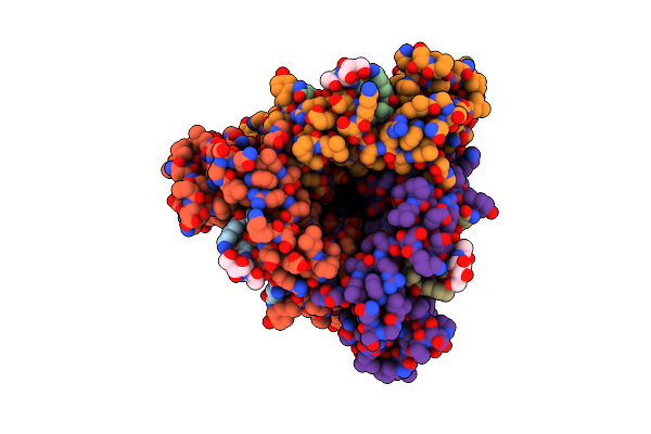
Organism: Norwalk virus
Method: ELECTRON MICROSCOPY Release Date: 2025-10-29 Classification: VIRAL PROTEIN Ligands: AGS, MG |
 |
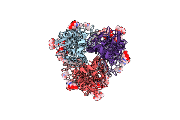
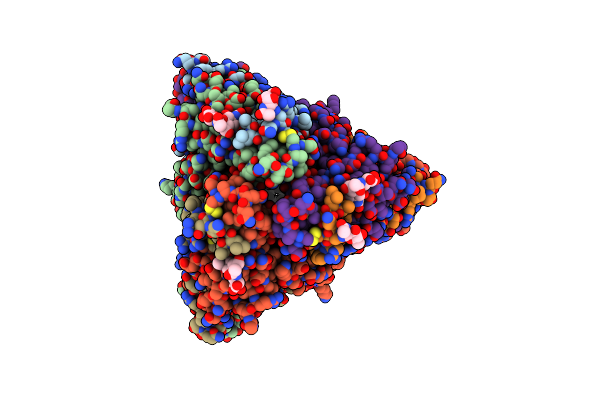
Organism: Influenza a virus, Aequorea victoria
Method: ELECTRON MICROSCOPY Release Date: 2025-08-13 Classification: VIRAL PROTEIN Ligands: NAG |
 |
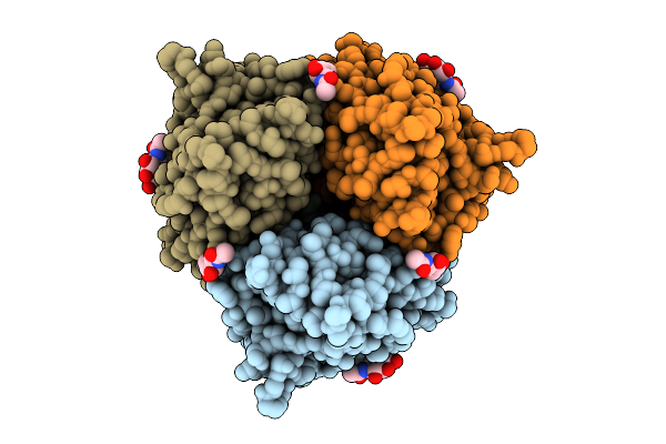
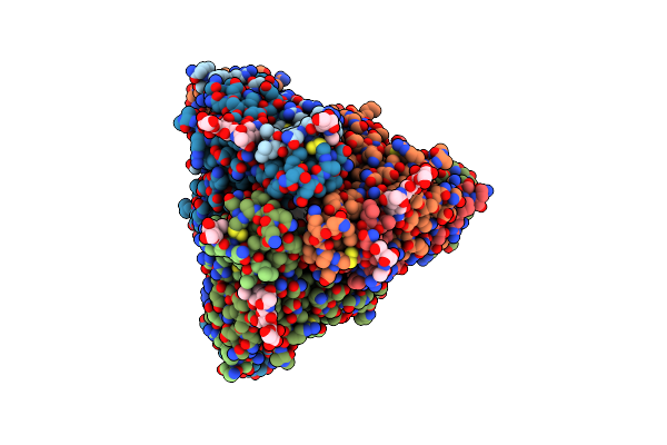
Organism: Influenza a virus
Method: ELECTRON MICROSCOPY Release Date: 2025-08-13 Classification: VIRAL PROTEIN |
 |
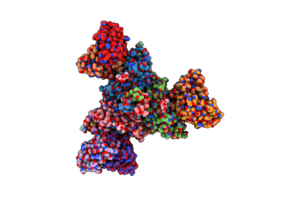
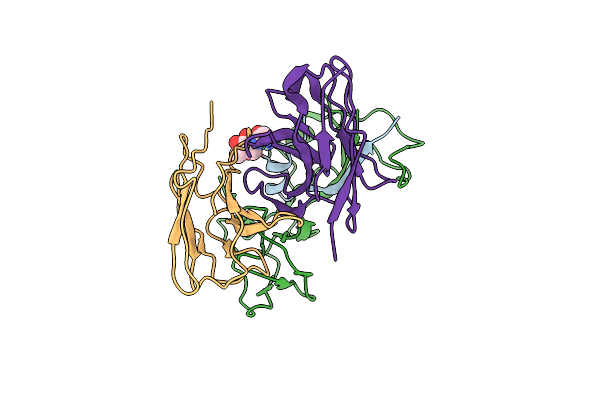
Organism: Influenza a virus
Method: ELECTRON MICROSCOPY Release Date: 2025-08-13 Classification: VIRAL PROTEIN Ligands: NAG |
 |
Organism: Influenza a virus (strain a/hong kong/1/1968 h3n2), Aequorea victoria
Method: ELECTRON MICROSCOPY Release Date: 2025-08-06 Classification: VIRAL PROTEIN Ligands: NAG |
 |
Organism: Influenza a virus (strain a/hong kong/1/1968 h3n2), Aequorea victoria
Method: ELECTRON MICROSCOPY Release Date: 2025-08-06 Classification: VIRAL PROTEIN Ligands: NAG |
 |
Respiratory Syncytial Virus Fusion Protein In The Postfusion Conformation In Complex With Monoclonal Antibody 131-2A Fab
Organism: Human respiratory syncytial virus, Mus musculus
Method: ELECTRON MICROSCOPY Release Date: 2025-02-26 Classification: VIRAL PROTEIN |
 |
Organism: Measles morbillivirus, Mus musculus
Method: ELECTRON MICROSCOPY Release Date: 2024-07-03 Classification: VIRAL PROTEIN Ligands: NAG |
 |
Structure Of The Measles Virus Fusion Protein In The Post-Fusion Conformation
Organism: Measles virus strain ichinose-b95a
Method: ELECTRON MICROSCOPY Release Date: 2024-07-03 Classification: VIRAL PROTEIN Ligands: NAG |
 |
Structure Of The Measles Virus Fusion Protein In The Pre-Fusion Conformation
Organism: Measles virus strain ichinose-b95a
Method: ELECTRON MICROSCOPY Release Date: 2024-07-03 Classification: VIRAL PROTEIN Ligands: NAG |
 |
Structure Of The Measles Virus Fusion Protein In The Pre-Fusion Conformation With Bound [Fip-Hrc]2-Peg11
Organism: Measles virus strain ichinose-b95a, Synthetic construct
Method: ELECTRON MICROSCOPY Release Date: 2024-07-03 Classification: VIRAL PROTEIN Ligands: NAG |
 |
Organism: Measles virus strain ichinose-b95a, Homo sapiens
Method: ELECTRON MICROSCOPY Release Date: 2024-07-03 Classification: VIRAL PROTEIN Ligands: NAG |
 |
Organism: Homo sapiens
Method: X-RAY DIFFRACTION Resolution:2.50 Å Release Date: 2024-04-03 Classification: IMMUNE SYSTEM |
 |
Crystal Structure Of Anti-Flag M2 Fab Fragment Bound To Flag-Tag Peptide Epitope
Organism: Mus musculus, Synthetic construct
Method: X-RAY DIFFRACTION Resolution:1.16 Å Release Date: 2024-04-03 Classification: IMMUNE SYSTEM Ligands: CL |
 |
Organism: Mus musculus
Method: X-RAY DIFFRACTION Resolution:3.50 Å Release Date: 2023-11-01 Classification: CELL ADHESION Ligands: NAG |
 |
Organism: Human coronavirus hku1
Method: ELECTRON MICROSCOPY Release Date: 2023-08-02 Classification: VIRAL PROTEIN Ligands: NAG |
 |
Human Coronavirus Hku1 Spike Glycoprotein In Complex With An Alpha2,8-Linked 9-O-Acetylated Disialoside (Closed State)
Organism: Human coronavirus hku1, Saccharomyces cerevisiae
Method: ELECTRON MICROSCOPY Release Date: 2023-08-02 Classification: VIRAL PROTEIN Ligands: NAG |
 |
Human Coronavirus Hku1 Spike Glycoprotein In Complex With An Alpha2,8-Linked 9-O-Acetylated Disialoside (1-Up State)
Organism: Human coronavirus hku1, Saccharomyces cerevisiae
Method: ELECTRON MICROSCOPY Release Date: 2023-08-02 Classification: VIRAL PROTEIN Ligands: NAG |
 |
Human Coronavirus Hku1 Spike Glycoprotein In Complex With An Alpha2,8-Linked 9-O-Acetylated Disialoside (3-Up State)
Organism: Human coronavirus hku1, Saccharomyces cerevisiae
Method: ELECTRON MICROSCOPY Release Date: 2023-08-02 Classification: VIRAL PROTEIN Ligands: NAG |
 |
Structure Of The Sars-Cov-2 Spike Glycoprotein In Complex With The Macrocyclic Peptide S1B3Inl1
Organism: Severe acute respiratory syndrome coronavirus 2, Tequatrovirus t4, Synthetic construct
Method: ELECTRON MICROSCOPY Release Date: 2023-06-28 Classification: VIRAL PROTEIN Ligands: NAG |