
Search Count: 36
 |
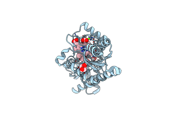
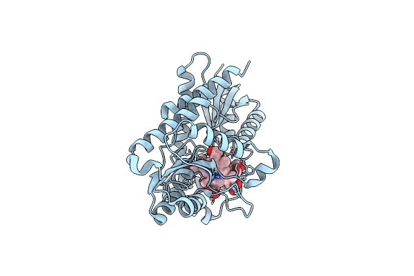
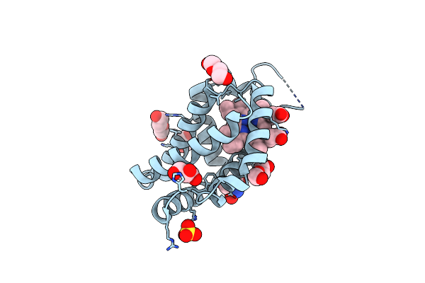
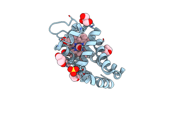
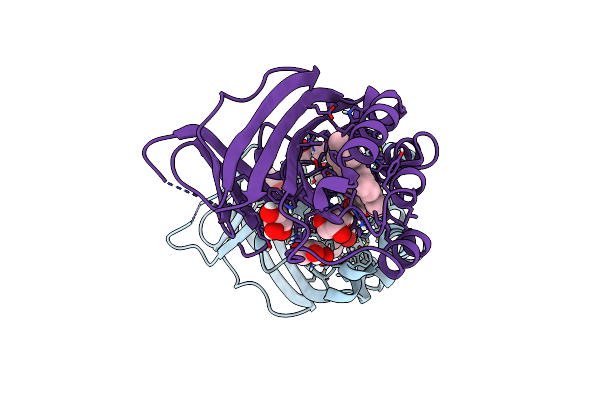
Coproporphyrin Iii - Lmcpfc Wt Complex Soaked With Fe2+ And Anomalous Densities
Organism: Listeria monocytogenes
Method: X-RAY DIFFRACTION Resolution:2.10 Å Release Date: 2024-04-24 Classification: METAL BINDING PROTEIN Ligands: HT9, EDO, FE, CL |
 |
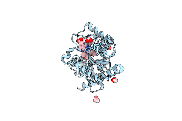
Organism: Listeria monocytogenes
Method: X-RAY DIFFRACTION Resolution:1.85 Å Release Date: 2024-04-24 Classification: METAL BINDING PROTEIN Ligands: FEC, EDO, CA |
 |
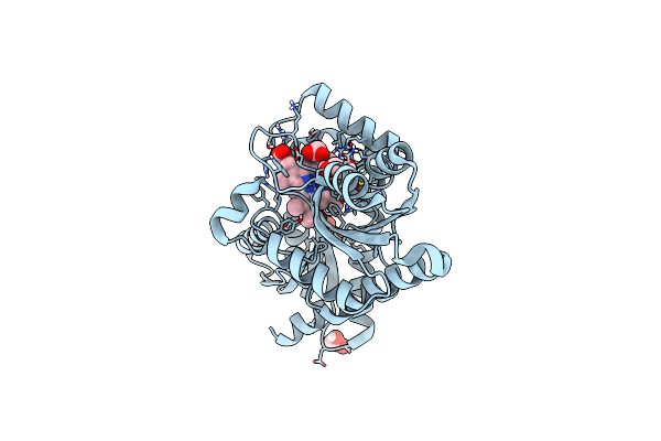
Organism: Listeria monocytogenes
Method: X-RAY DIFFRACTION Resolution:2.15 Å Release Date: 2023-05-03 Classification: METAL BINDING PROTEIN Ligands: GOL, EDO, HT9, FE2, MG |
 |
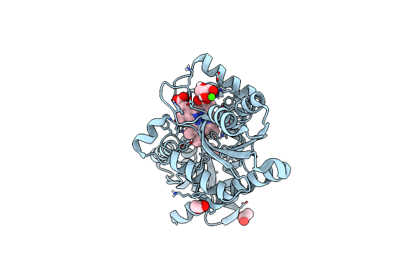
Organism: Listeria monocytogenes
Method: X-RAY DIFFRACTION Resolution:2.10 Å Release Date: 2023-04-05 Classification: METAL BINDING PROTEIN Ligands: GOL, ACT, HT9, EDO, FE2 |
 |
Organism: Listeria monocytogenes
Method: X-RAY DIFFRACTION Resolution:2.19 Å Release Date: 2023-01-11 Classification: METAL BINDING PROTEIN Ligands: GOL, EDO, HT9, FE2, CL |
 |
Organism: Listeria monocytogenes
Method: X-RAY DIFFRACTION Resolution:1.51 Å Release Date: 2022-12-28 Classification: METAL BINDING PROTEIN Ligands: ACT, GOL, EDO, HT9, CA |
 |
Organism: Listeria monocytogenes
Method: X-RAY DIFFRACTION Resolution:2.64 Å Release Date: 2022-12-28 Classification: METAL BINDING PROTEIN Ligands: GOL, HT9 |
 |
Organism: Mus musculus
Method: X-RAY DIFFRACTION Resolution:1.80 Å Release Date: 2022-07-13 Classification: OXYGEN TRANSPORT Ligands: HEM, SO4, GOL, IPA, BTB |
 |
M.Tuberculosis Nitrobindin With A Water Molecule Coordinated To The Heme Iron Atom
Organism: Mycobacterium tuberculosis
Method: X-RAY DIFFRACTION Resolution:1.20 Å Release Date: 2020-04-08 Classification: PROTEIN BINDING Ligands: HEM |
 |
M.Tuberculosis Nitrobindin With A Cyanide Molecule Coordinated To The Heme Iron Atom
Organism: Mycobacterium tuberculosis
Method: X-RAY DIFFRACTION Resolution:1.60 Å Release Date: 2020-04-08 Classification: PROTEIN BINDING Ligands: HEM, CYN |
 |
Organism: Mus musculus
Method: X-RAY DIFFRACTION Resolution:2.30 Å Release Date: 2020-03-11 Classification: OXYGEN BINDING Ligands: HEM, SO4, TRS, PEG, GOL |
 |
Structure Of Coproheme Decarboxylase From Listeria Monocytogenes In Complex With Iron Coproporphyrin Iii
Organism: Listeria monocytogenes egd-e
Method: X-RAY DIFFRACTION Release Date: 2019-07-10 Classification: OXIDOREDUCTASE |
 |
Structure Of Coproheme Decarboxylase From Listeria Monocytogenes During Turnover
Organism: Listeria monocytogenes serovar 1/2a (strain atcc baa-679 / egd-e)
Method: X-RAY DIFFRACTION Resolution:1.69 Å Release Date: 2019-07-10 Classification: OXIDOREDUCTASE |
 |
Organism: Mus musculus
Method: X-RAY DIFFRACTION Resolution:1.80 Å Release Date: 2019-04-10 Classification: OXYGEN BINDING Ligands: HEM, SO4, DIO |
 |
Organism: Mus musculus
Method: X-RAY DIFFRACTION Resolution:1.75 Å Release Date: 2019-04-10 Classification: OXYGEN BINDING Ligands: HEM, CMO, SO4, DIO, GOL |
 |
Organism: Mus musculus
Method: X-RAY DIFFRACTION Resolution:1.60 Å Release Date: 2019-04-10 Classification: OXYGEN BINDING Ligands: HEM, SO4, TRS, GOL, PEG |
 |
Organism: Mus musculus
Method: X-RAY DIFFRACTION Resolution:2.60 Å Release Date: 2019-04-10 Classification: OXYGEN BINDING Ligands: HEM, CMO, SO4, TRS, GOL, PEG |
 |
Joint Neutron/X-Ray Structure Of Dimeric Chlorite Dismutase From Cyanothece Sp. Pcc7425
Organism: Cyanothece sp. (strain pcc 7425 / atcc 29141)
Method: X-RAY DIFFRACTION, NEUTRON DIFFRACTION Resolution:2.000 Å, 2.350 Å Release Date: 2018-02-28 Classification: OXIDOREDUCTASE Ligands: HEM, OH, CL, GOL |
 |
Crystal Structure Of Dimeric Chlorite Dismutase From Cyanothece Sp. Pcc7425 At Ph 9.0 And 293 K.
Organism: Cyanothece sp. pcc 7425
Method: X-RAY DIFFRACTION Resolution:2.00 Å Release Date: 2018-01-31 Classification: OXIDOREDUCTASE Ligands: GOL, HEM, CL |
 |
Crystal Structure Of Dimeric Chlorite Dismutase From Cyanothece Sp. Pcc7425 (Ph 6.5)
Organism: Cyanothece sp. pcc 7425
Method: X-RAY DIFFRACTION Resolution:1.30 Å Release Date: 2017-12-20 Classification: OXIDOREDUCTASE Ligands: HEM, SO4, GOL |