
Search Count: 42
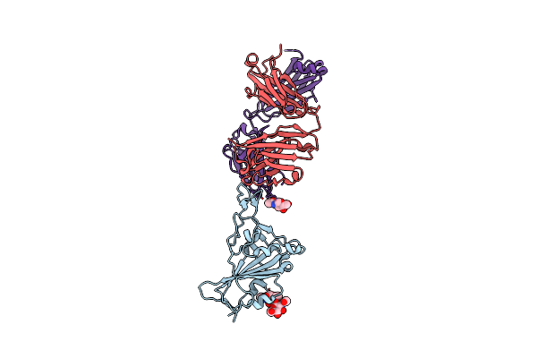 |
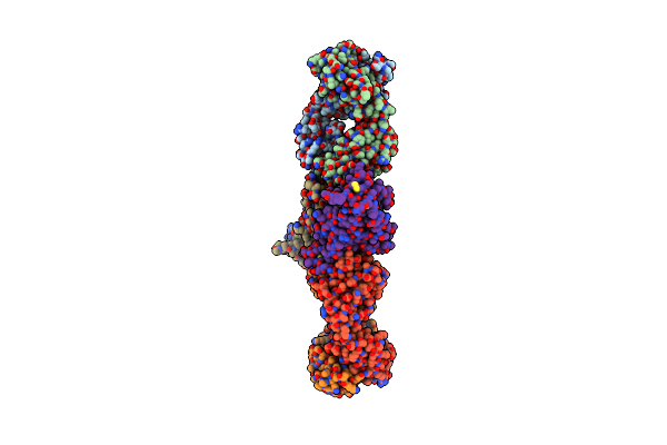
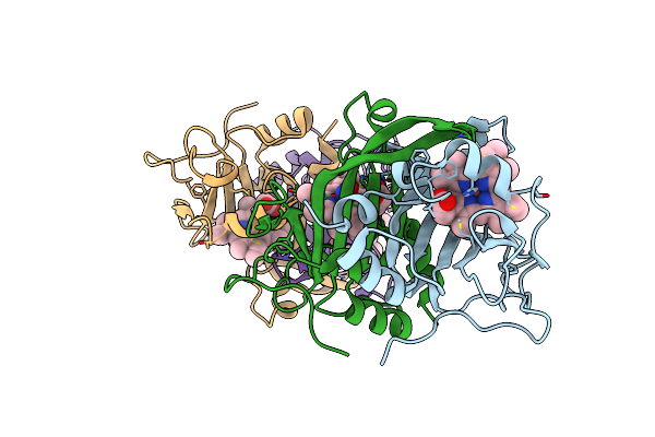
Crystal Structure Of Sars-Cov-2 Rbd In Complex With The Ridge-Binding Nab Eh8 Isolated From A Nonvaccinated Pediatric Patient
Organism: Severe acute respiratory syndrome coronavirus 2, Homo sapiens
Method: X-RAY DIFFRACTION Resolution:2.49 Å Release Date: 2023-03-08 Classification: VIRAL PROTEIN/Immune System Ligands: NAG, NA |
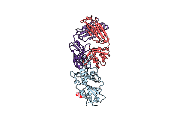 |
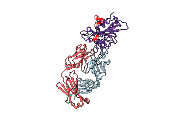
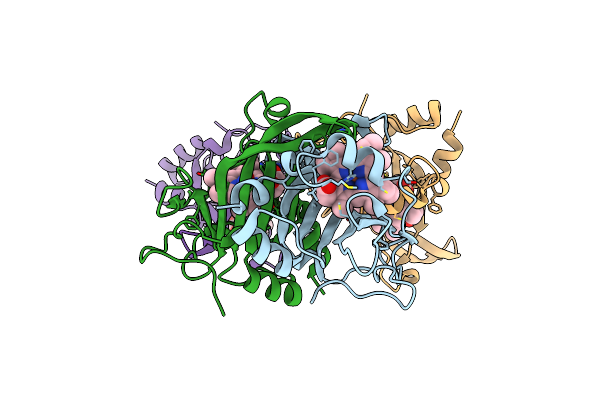
Crystal Structure Of Sars-Cov-2 Rbd In Complex With The Neutralizing Ighv3-53-Encoded Antibody Eh3 Isolated From A Nonvaccinated Pediatric Patient
Organism: Severe acute respiratory syndrome coronavirus, Homo sapiens
Method: X-RAY DIFFRACTION Resolution:2.65 Å Release Date: 2023-03-08 Classification: VIRAL PROTEIN/Immune System Ligands: NAG |
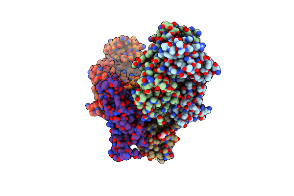 |
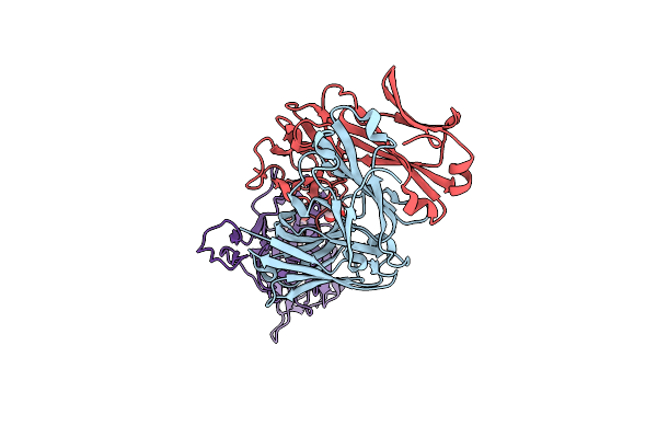
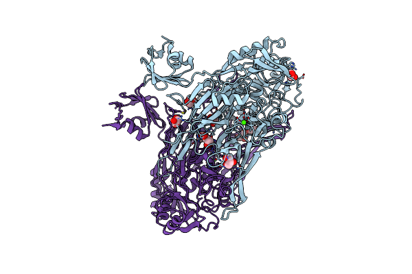
Organism: Homo sapiens, Severe acute respiratory syndrome coronavirus 2
Method: X-RAY DIFFRACTION Resolution:2.43 Å Release Date: 2021-05-12 Classification: IMMUNE SYSTEM/VIRAL PROTEIN Ligands: NAG |
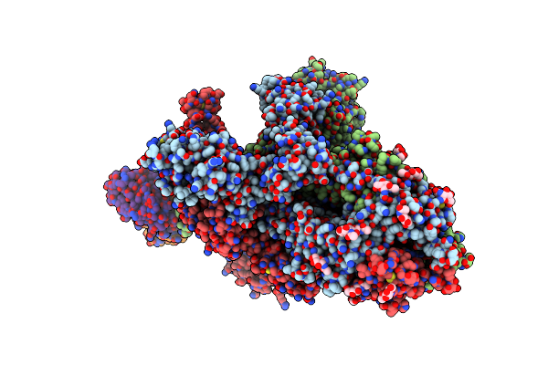 |
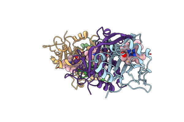
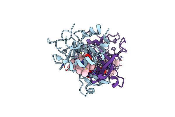
Organism: Severe acute respiratory syndrome coronavirus 2, Homo sapiens
Method: ELECTRON MICROSCOPY Release Date: 2021-02-03 Classification: VIRAL PROTEIN/IMMUNE SYSTEM Ligands: NAG |
 |
Organism: Homo sapiens, Severe acute respiratory syndrome coronavirus 2
Method: X-RAY DIFFRACTION Resolution:2.16 Å Release Date: 2021-01-27 Classification: IMMUNE SYSTEM/VIRAL PROTEIN Ligands: GOL |
 |
Organism: Homo sapiens, Severe acute respiratory syndrome coronavirus 2
Method: X-RAY DIFFRACTION Resolution:1.72 Å Release Date: 2021-01-27 Classification: IMMUNE SYSTEM/VIRAL PROTEIN Ligands: NAG, GOL, PRO |
 |
Organism: Homo sapiens, Severe acute respiratory syndrome coronavirus 2
Method: X-RAY DIFFRACTION Resolution:1.73 Å Release Date: 2021-01-27 Classification: IMMUNE SYSTEM/VIRAL PROTEIN Ligands: GOL, NAG |
 |
Organism: Homo sapiens
Method: X-RAY DIFFRACTION Resolution:1.90 Å Release Date: 2019-12-18 Classification: TRANSCRIPTION Ligands: OP4 |
 |
Organism: Nitrosomonas sp. al212
Method: X-RAY DIFFRACTION Resolution:1.97 Å Release Date: 2019-02-27 Classification: METAL BINDING PROTEIN Ligands: HEC |
 |
Organism: Nitrosomonas sp. al212
Method: X-RAY DIFFRACTION Resolution:2.26 Å Release Date: 2019-02-27 Classification: METAL BINDING PROTEIN Ligands: HEC, HOA |
 |
Organism: Nitrosomonas sp. al212
Method: X-RAY DIFFRACTION Resolution:2.30 Å Release Date: 2019-02-27 Classification: METAL BINDING PROTEIN Ligands: HEC |
 |
Organism: Nitrosomonas sp. al212
Method: X-RAY DIFFRACTION Resolution:1.97 Å Release Date: 2019-02-27 Classification: METAL BINDING PROTEIN Ligands: HEC, NO |
 |
Organism: Escherichia coli
Method: X-RAY DIFFRACTION Resolution:1.70 Å Release Date: 2018-09-05 Classification: OXIDOREDUCTASE Ligands: CU, CA, GOL |
 |
Organism: Escherichia coli (strain k12)
Method: X-RAY DIFFRACTION Resolution:1.80 Å Release Date: 2018-08-29 Classification: OXIDOREDUCTASE Ligands: CU, CA, GOL |
 |
Organism: Nitrosomonas sp. al212
Method: X-RAY DIFFRACTION Resolution:1.45 Å Release Date: 2017-12-20 Classification: METAL BINDING PROTEIN Ligands: HEC |
 |
Organism: Schizosaccharomyces pombe
Method: X-RAY DIFFRACTION Resolution:2.75 Å Release Date: 2017-02-08 Classification: RECOMBINATION Ligands: SO4, GOL |
 |
Structural And Biochemical Studies Of A Moderately Thermophilic Exonuclease I From Methylocaldum Szegediense
Organism: Methylocaldum szegediense
Method: X-RAY DIFFRACTION Resolution:2.12 Å Release Date: 2015-02-25 Classification: HYDROLASE Ligands: MG |
 |
Structure Of Keap1 Kelch Domain With (1S,2R)-2-{[(1S)-1-[(1,3-Dioxo-1,3-Dihydro-2H-Isoindol-2-Yl)Methyl]-3,4-Dihydroisoquinolin-2(1H)-Yl]Carbonyl}Cyclohexanecarboxylic Acid
Organism: Homo sapiens
Method: X-RAY DIFFRACTION Resolution:2.41 Å Release Date: 2014-02-19 Classification: TRANSCRIPTION/INHIBITOR Ligands: ACT, 1VV, NA |
 |
Structure Of Keap1 Kelch Domain With 2-{[(1S)-2-{[(1R,2S)-2-(1H-Tetrazol-5-Yl)Cyclohexyl]Carbonyl}-1,2,3,4-Tetrahydroisoquinolin-1-Yl]Methyl}-1H-Isoindole-1,3(2H)-Dione
Organism: Homo sapiens
Method: X-RAY DIFFRACTION Resolution:2.40 Å Release Date: 2014-02-19 Classification: TRANSCRIPTION/INHIBITOR Ligands: 1VW, ACT |
 |
Structure Of Keap1 Kelch Domain With (1S,2R)-2-{[(1S)-5-Methyl-1-[(1-Oxo-1,3-Dihydro-2H-Isoindol-2-Yl)Methyl]-3,4-Dihydroisoquinolin-2(1H)-Yl]Carbonyl}Cyclohexanecarboxylic Acid
Organism: Homo sapiens
Method: X-RAY DIFFRACTION Resolution:2.25 Å Release Date: 2014-02-19 Classification: TRANSCRIPTION/INHIBITOR Ligands: 1VX, ACT |