
Search Count: 9
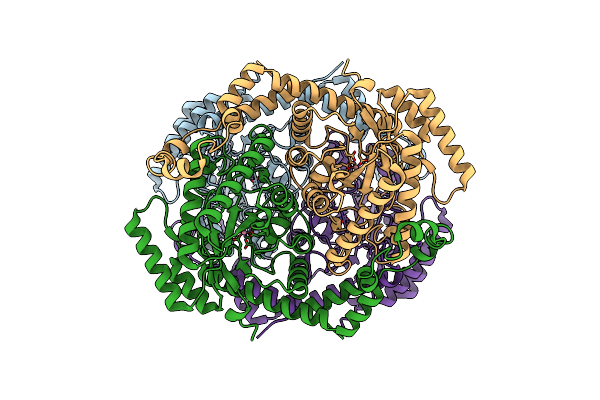 |
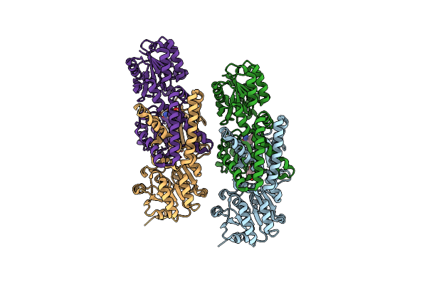
Cryo-Electron Microscopy Structure Of Glucose/Xylose Isomerase From Streptomyces Rubiginosus With Cobalt Ions In The Active Site
Organism: Streptomyces rubiginosus
Method: ELECTRON MICROSCOPY Release Date: 2024-10-02 Classification: METAL BINDING PROTEIN Ligands: CO |
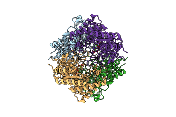 |
Cryo-Electron Microscopy Structure Of Glucose/Xylose Isomerase From Streptomyces Rubiginosus With Magnesium Ions In The Active Site
Organism: Streptomyces rubiginosus
Method: ELECTRON MICROSCOPY Release Date: 2024-10-02 Classification: METAL BINDING PROTEIN Ligands: MG |
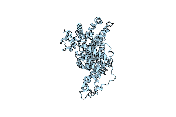 |
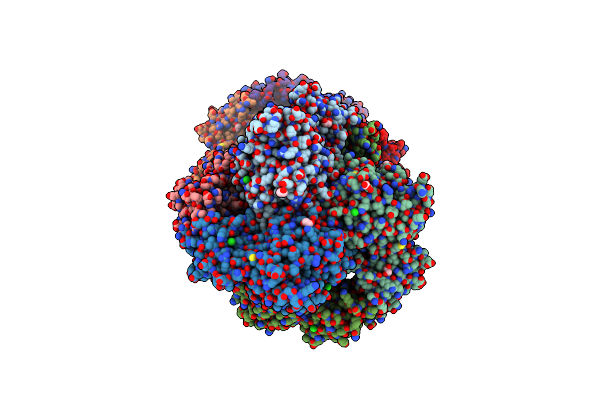
Structural Studies Of Human Serum Albumin Using Cryo-Em Up To 0.38 Nm Resolution
Organism: Homo sapiens
Method: ELECTRON MICROSCOPY Release Date: 2023-08-16 Classification: TRANSPORT PROTEIN |
 |
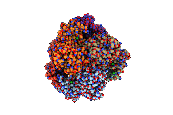
Crystal Structure Of Prephenate Dehydrogenase Tyra From Bacillus Anthracis In Complex With Nad And L-Tyrosine
Organism: Bacillus anthracis
Method: X-RAY DIFFRACTION Resolution:2.10 Å Release Date: 2019-09-11 Classification: HYDROLASE Ligands: TYR, PO4, NAD |
 |
2.6 Angstrom Resolution Crystal Structure Of Aminopeptidase B From Escherichia Coli Str. K-12 Substr. Mg1655
Organism: Escherichia coli (strain k12)
Method: X-RAY DIFFRACTION Resolution:2.61 Å Release Date: 2019-05-15 Classification: HYDROLASE Ligands: ZN, MN, CL |
 |
2.05 Angstrom Resolution Crystal Structure Of Aminopeptidase B From Escherichia Coli Str. K-12 Substr. Mg1655.
Organism: Escherichia coli str. k-12 substr. mg1655
Method: X-RAY DIFFRACTION Resolution:2.05 Å Release Date: 2019-03-27 Classification: HYDROLASE Ligands: ZN, CA, CL, BCT, EDO, PGE, PEG |
 |
Crystal Structure Of Peptidase B From Yersinia Pestis Co92 At 2.75 A Resolution
Organism: Yersinia pestis co92
Method: X-RAY DIFFRACTION Resolution:2.75 Å Release Date: 2018-04-18 Classification: HYDROLASE Ligands: SO4 |
 |
Crystal Structure Of Prephenate Dehydrogenase Tyra From Bacillus Anthracis In Complex With L-Tyrosine
Organism: Bacillus anthracis
Method: X-RAY DIFFRACTION Resolution:2.60 Å Release Date: 2017-03-08 Classification: OXIDOREDUCTASE Ligands: TYR |
 |
Crystal Structure Of The Act Domain Of Prephenate Dehydrogenase Tyra From Bacillus Anthracis
Organism: Bacillus anthracis
Method: X-RAY DIFFRACTION Resolution:2.01 Å Release Date: 2017-03-08 Classification: OXIDOREDUCTASE Ligands: SO4, CA |