
Search Count: 26
 |
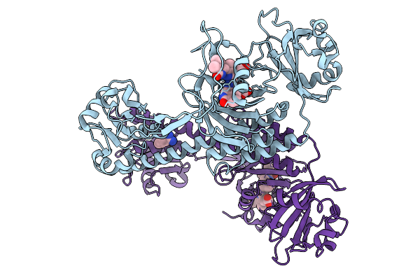
Photoactivation In Bacteriophytochromes, Reference (Dark) Structure For The 3 Ps Time Point
Organism: Stigmatella aurantiaca
Method: X-RAY DIFFRACTION Release Date: 2025-10-08 Classification: SIGNALING PROTEIN Ligands: 3Q8, BEN |
 |
Photoactivation In Bacteriophytochrome, High Resolution Cryo Structure In The Dark.
Organism: Stigmatella aurantiaca
Method: X-RAY DIFFRACTION Release Date: 2025-10-08 Classification: SIGNALING PROTEIN Ligands: EL5, P33 |
 |
Photoactivation In Bacteriophytochromes, Reference (Dark) Structure For The 100 Ps Time Point
Organism: Stigmatella aurantiaca
Method: X-RAY DIFFRACTION Release Date: 2025-10-08 Classification: SIGNALING PROTEIN Ligands: EL5, BEN |
 |
Organism: Stigmatella aurantiaca
Method: X-RAY DIFFRACTION Release Date: 2025-10-08 Classification: SIGNALING PROTEIN Ligands: BLA, BEN |
 |
Organism: Stigmatella aurantiaca
Method: X-RAY DIFFRACTION Release Date: 2025-10-08 Classification: SIGNALING PROTEIN Ligands: BLA, BEN |
 |
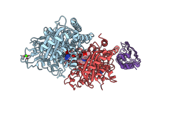
Dark Structure Of The Human Adenosine A2A Receptor Bound To Synthetic Photoswitch "Stilswitch3" Determined By Serial Synchrotron Crystallography
Organism: Homo sapiens
Method: X-RAY DIFFRACTION Resolution:2.65 Å Release Date: 2025-01-08 Classification: MEMBRANE PROTEIN Ligands: OLA, A1H3H, CLR, NA |
 |
Crystal Structure Of The Adenosine A2A Receptor In Complex With Istradefylline
Organism: Homo sapiens
Method: X-RAY DIFFRACTION Resolution:1.94 Å Release Date: 2025-01-08 Classification: MEMBRANE PROTEIN Ligands: JQ9, CLR, OLA, NA |
 |
Crystal Structure Of The Adenosine A2A Receptor In Complex With The Synthetic Photoswitch 'Azoswitch2
Organism: Homo sapiens
Method: X-RAY DIFFRACTION Resolution:2.20 Å Release Date: 2025-01-08 Classification: MEMBRANE PROTEIN Ligands: A1H3L, CLR, OLA, NA |
 |
Crystal Structure Of The Adenosine A2A Receptor In Complex With The Synthetic Photoswitch 'Stilswitch1
Organism: Homo sapiens
Method: X-RAY DIFFRACTION Resolution:2.25 Å Release Date: 2025-01-08 Classification: MEMBRANE PROTEIN Ligands: A1H3J, CLR, OLA, OLC, OLB, NA |
 |
Crystal Structure Of The Adenosine A2A Receptor In Complex With The Synthetic Photoswitch 'Stilswitch2
Organism: Homo sapiens
Method: X-RAY DIFFRACTION Resolution:2.31 Å Release Date: 2025-01-08 Classification: MEMBRANE PROTEIN Ligands: A1H3I, CLR, OLC, OLA, NA |
 |
Crystal Structure Of The Adenosine A2A Receptor In Complex With The Synthetic Photoswitch 'Stilswitch3
Organism: Homo sapiens
Method: X-RAY DIFFRACTION Resolution:2.05 Å Release Date: 2025-01-08 Classification: MEMBRANE PROTEIN Ligands: A1H3H, CLR, OLA, NA |
 |
Crystal Structure Of The Adenosine A2A Receptor In Complex With The Synthetic Photoswitch 'Stilswitch4
Organism: Homo sapiens
Method: X-RAY DIFFRACTION Resolution:2.20 Å Release Date: 2025-01-08 Classification: MEMBRANE PROTEIN Ligands: A1H3K, CLR, OLA, OLC, NA |
 |
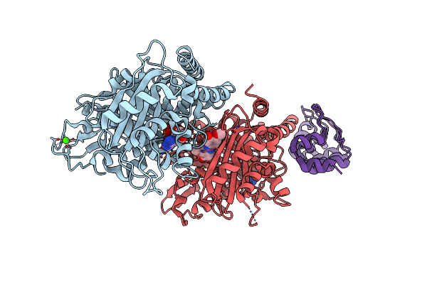
Dark Structure Of The Human Adenosine A2A Receptor Bound To Synthetic Photoswitch 'Stilswitch2' Determined By Serial Synchrotron Crystallography
Organism: Homo sapiens
Method: X-RAY DIFFRACTION Resolution:2.45 Å Release Date: 2025-01-08 Classification: MEMBRANE PROTEIN Ligands: A1H3I, CLR, OLA, OLB, OLC, NA |
 |
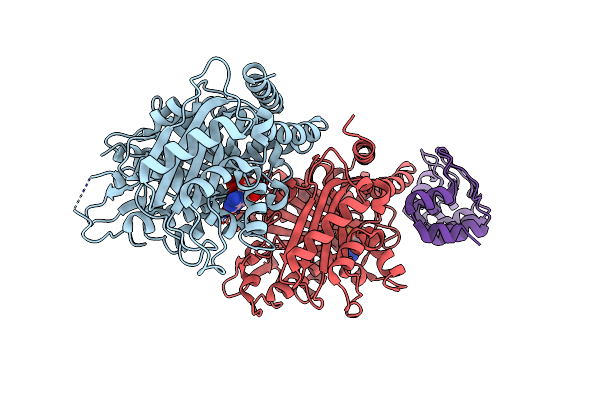
Steady State Structure Of The Human Adenosine A2A Receptor Bound To Synthetic Photoswitch 'Stilswitch2' Determined By Serial Synchrotron Crystallography
Organism: Homo sapiens
Method: X-RAY DIFFRACTION Resolution:2.80 Å Release Date: 2025-01-08 Classification: MEMBRANE PROTEIN Ligands: A1H3I, CLR, OLA, NA |
 |
Steady State Structure Of The Human Adenosine A2A Receptor Bound To Synthetic Photoswitch 'Stilswitch3' Determined By Serial Synchrotron Crystallography
Organism: Homo sapiens, Escherichia coli
Method: X-RAY DIFFRACTION Resolution:3.05 Å Release Date: 2025-01-08 Classification: MEMBRANE PROTEIN Ligands: A1H3H, OLA, NA |
 |
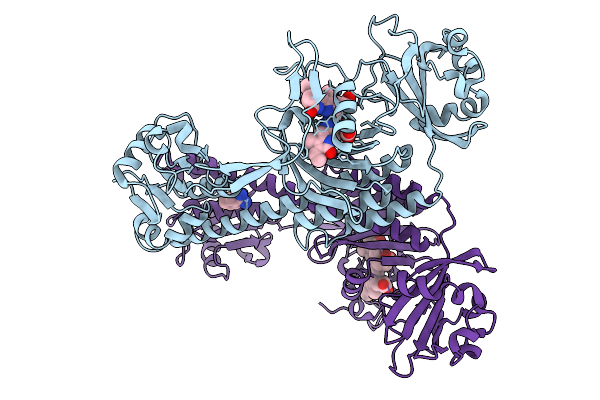
Molecular Snapshots Of Drug Release From Tubulin: 1 Microsecond After Photoactivation
Organism: Synthetic construct, Bos taurus
Method: X-RAY DIFFRACTION Resolution:2.20 Å Release Date: 2023-02-22 Classification: CELL CYCLE Ligands: GTP, MG, CA, GDP, VYT |
 |
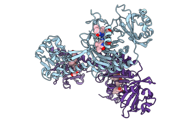
Molecular Snapshots Of Drug Release From Tubulin: 10 Microseconds After Photoactivation.
Organism: Synthetic construct, Bos taurus
Method: X-RAY DIFFRACTION Resolution:2.20 Å Release Date: 2023-02-22 Classification: CELL CYCLE Ligands: GTP, MG, CA, GDP, VYT |
 |
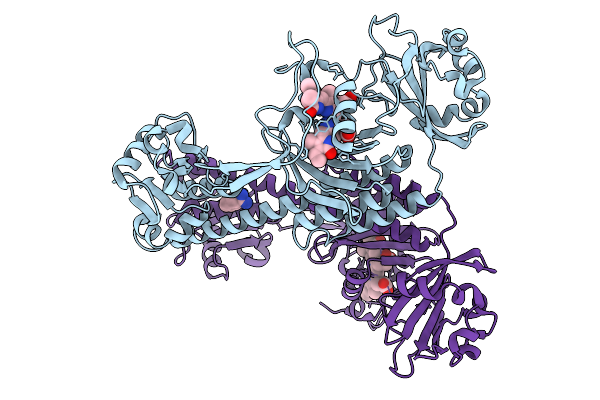
Molecular Snapshots Of Drug Release From Tubulin: 100 Microseconds After Photoactivation.
Organism: Synthetic construct, Bos taurus
Method: X-RAY DIFFRACTION Resolution:2.20 Å Release Date: 2023-02-22 Classification: CELL CYCLE Ligands: GTP, MG, CA, GDP, VYT |
 |
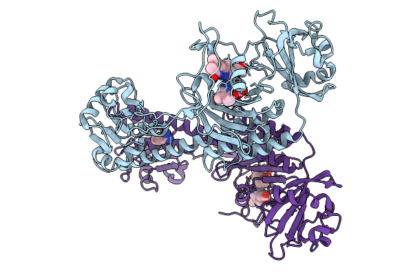
Molecular Snapshots Of Drug Release From Tubulin: 1 Millisecond After Photoactivation.
Organism: Synthetic construct, Bos taurus
Method: X-RAY DIFFRACTION Resolution:2.20 Å Release Date: 2023-02-22 Classification: CELL CYCLE Ligands: GTP, MG, CA, GDP, VYT |
 |
Molecular Snapshots Of Drug Release From Tubulin: 10 Milliseconds After Photoactivation.
Organism: Synthetic construct, Bos taurus
Method: X-RAY DIFFRACTION Resolution:2.20 Å Release Date: 2023-02-22 Classification: CELL CYCLE Ligands: GTP, MG, GDP |