
Search Count: 14
 |
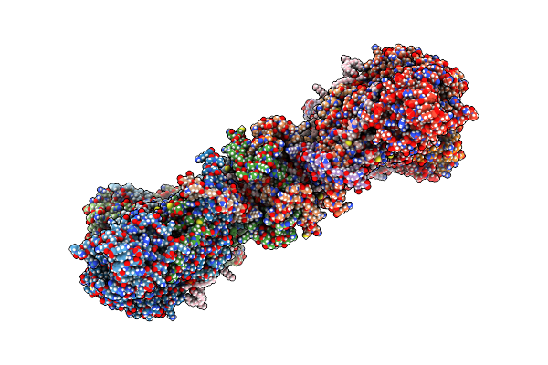
Organism: Trypanosoma brucei brucei
Method: ELECTRON MICROSCOPY Release Date: 2022-12-07 Classification: MEMBRANE PROTEIN Ligands: ATP, MG, CDL, ADP, LMT, Q7G, PEE, PC1, UTP |
 |
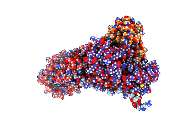
Organism: Trypanosoma brucei brucei
Method: ELECTRON MICROSCOPY Release Date: 2022-10-26 Classification: MEMBRANE PROTEIN Ligands: CDL, LMT, Q7G, PEE, PC1 |
 |
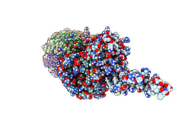
Organism: Trypanosoma brucei brucei
Method: ELECTRON MICROSCOPY Release Date: 2022-10-26 Classification: MEMBRANE PROTEIN |
 |
Organism: Trypanosoma brucei brucei
Method: ELECTRON MICROSCOPY Release Date: 2022-10-26 Classification: MEMBRANE PROTEIN Ligands: UTP |
 |
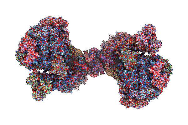
Rotational State 1A Of The Trypanosoma Brucei Mitochondrial Atp Synthase Dimer
Organism: Trypanosoma brucei brucei
Method: ELECTRON MICROSCOPY Release Date: 2022-10-26 Classification: MEMBRANE PROTEIN Ligands: ATP, MG, ADP, UTP, CDL, PEE, LMT, Q7G, PC1 |
 |
Rotational State 1B Of The Trypanosoma Brucei Mitochondrial Atp Synthase Dimer
Organism: Trypanosoma brucei brucei
Method: ELECTRON MICROSCOPY Release Date: 2022-10-26 Classification: MEMBRANE PROTEIN Ligands: ATP, MG, ADP, UTP, CDL, 3PE, PC1, LMT, Q7G |
 |
Rotational State 1C Of The Trypanosoma Brucei Mitochondrial Atp Synthase Dimer
Organism: Trypanosoma brucei brucei
Method: ELECTRON MICROSCOPY Release Date: 2022-10-26 Classification: MEMBRANE PROTEIN Ligands: ATP, MG, ADP, UTP, CDL, 3PE, LMT, Q7G, PC1 |
 |
Rotational State 1D Of The Trypanosoma Brucei Mitochondrial Atp Synthase Dimer
Organism: Trypanosoma brucei brucei
Method: ELECTRON MICROSCOPY Release Date: 2022-10-26 Classification: MEMBRANE PROTEIN Ligands: CDL, PEE, PC1, LMT, Q7G, ATP, MG, ADP, UTP |
 |
Rotational State 1E Of The Trypanosoma Brucei Mitochondrial Atp Synthase Dimer
Organism: Trypanosoma brucei brucei
Method: ELECTRON MICROSCOPY Release Date: 2022-10-26 Classification: MEMBRANE PROTEIN Ligands: ATP, MG, ADP, UTP, CDL, PEE, PC1, LMT, Q7G |
 |
Rotational State 2A Of The Trypanosoma Brucei Mitochondrial Atp Synthase Dimer
Organism: Trypanosoma brucei brucei
Method: ELECTRON MICROSCOPY Release Date: 2022-10-26 Classification: MEMBRANE PROTEIN Ligands: CDL, PEE, LMT, Q7G, PC1, UTP, ATP, MG, ADP |
 |
Rotational State 2B Of The Trypanosoma Brucei Mitochondrial Atp Synthase Dimer
Organism: Trypanosoma brucei brucei
Method: ELECTRON MICROSCOPY Release Date: 2022-10-26 Classification: MEMBRANE PROTEIN Ligands: CDL, PEE, LMT, Q7G, PC1, UTP, ATP, MG, ADP |
 |
Rotational State 2C Of The Trypanosoma Brucei Mitochondrial Atp Synthase Dimer
Organism: Trypanosoma brucei brucei
Method: ELECTRON MICROSCOPY Release Date: 2022-10-26 Classification: MEMBRANE PROTEIN Ligands: CDL, PEE, LMT, Q7G, PC1, UTP, ATP, MG, ADP |
 |
Organism: Trypanosoma brucei brucei
Method: ELECTRON MICROSCOPY Release Date: 2022-10-26 Classification: MEMBRANE PROTEIN Ligands: CDL, PEE, LMT, Q7G, PC1, UTP, ATP, MG, ADP |
 |
Rotational State 3 Of The Trypanosoma Brucei Mitochondrial Atp Synthase Dimer
Organism: Trypanosoma brucei brucei
Method: ELECTRON MICROSCOPY Release Date: 2022-10-26 Classification: MEMBRANE PROTEIN Ligands: CDL, 3PE, PC1, LMT, Q7G, ATP, MG, ADP, UTP |