
Search Count: 31
 |
Organism: Mus musculus
Method: ELECTRON MICROSCOPY Release Date: 2025-08-06 Classification: MEMBRANE PROTEIN Ligands: MG, KXP, BEF, NAG |
 |
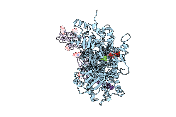
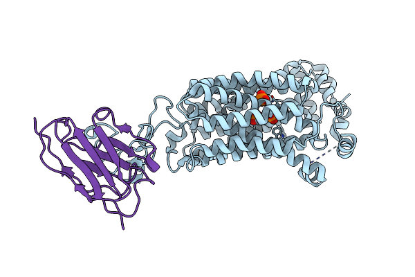
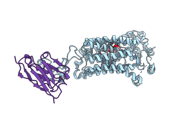
Cryo-Em Structure Of Mouse Pmca-Nptn Complex Captured In E1 State Without Calcium
Organism: Mus musculus
Method: ELECTRON MICROSCOPY Release Date: 2025-08-06 Classification: MEMBRANE PROTEIN Ligands: KXP, NAG |
 |
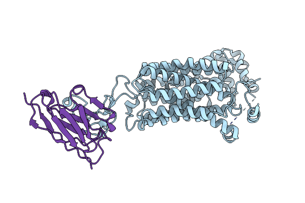
Organism: Mus musculus
Method: ELECTRON MICROSCOPY Release Date: 2025-08-06 Classification: MEMBRANE PROTEIN Ligands: ANP, MG, KXP, NAG |
 |
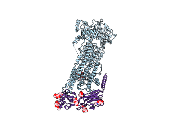
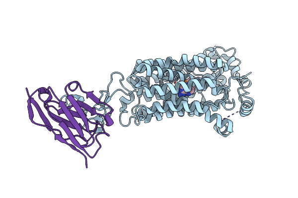
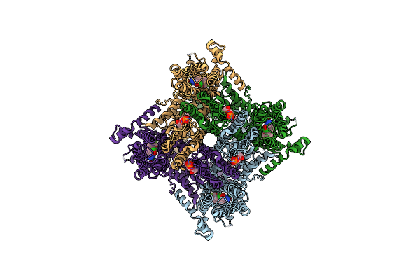
Cryo-Em Structure Of Mouse Pmca-Nptn Complex Captured In E2-Pi State (Alf4)
Organism: Mus musculus
Method: ELECTRON MICROSCOPY Release Date: 2025-08-06 Classification: MEMBRANE PROTEIN Ligands: ALF, MG, NAG |
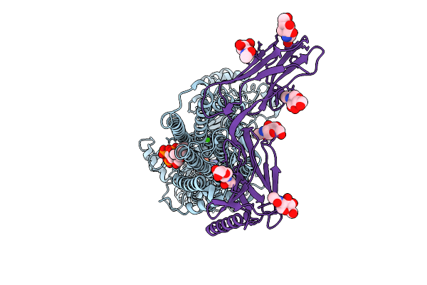 |
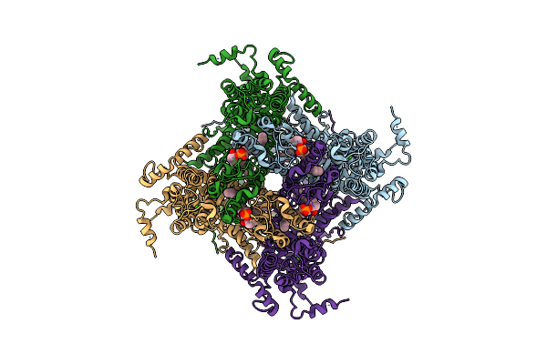
Organism: Mus musculus
Method: ELECTRON MICROSCOPY Release Date: 2025-08-06 Classification: MEMBRANE PROTEIN Ligands: ANP, CA, MG, KXP, NAG |
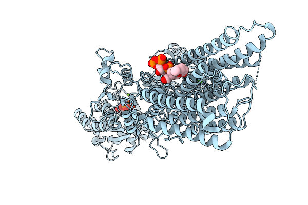 |
Cryo-Em Structure Of Mouse Pmca Captured In E1-Atp In The Presence Of Calcium
Organism: Mus musculus
Method: ELECTRON MICROSCOPY Release Date: 2025-08-06 Classification: MEMBRANE PROTEIN Ligands: ANP, CA, MG, KXP |
 |
Organism: Mus musculus
Method: ELECTRON MICROSCOPY Release Date: 2025-08-06 Classification: MEMBRANE PROTEIN Ligands: BEF, MG, KXP |
 |
Organism: Mus musculus
Method: ELECTRON MICROSCOPY Release Date: 2025-08-06 Classification: MEMBRANE PROTEIN Ligands: CA, KXP, NAG |
 |
Organism: Mus musculus
Method: X-RAY DIFFRACTION Release Date: 2025-08-06 Classification: CELL ADHESION Ligands: PEG, EPE |
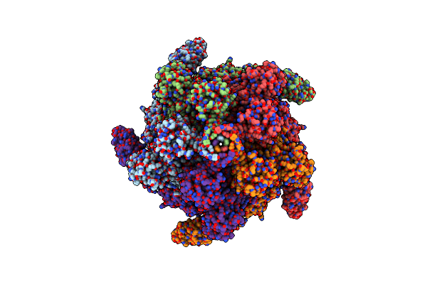 |
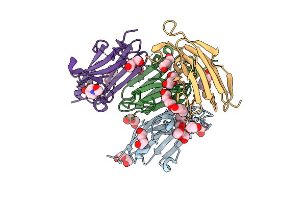
Organism: Photorhabdus luminescens
Method: ELECTRON MICROSCOPY Release Date: 2024-03-13 Classification: TOXIN |
 |
Photorhabdus Luminescens Tcda1 Prepore-To-Pore Intermediate, K567W K2008W Mutant
Organism: Photorhabdus luminescens
Method: ELECTRON MICROSCOPY Release Date: 2024-03-13 Classification: TOXIN |
 |
Photorhabdus Luminescens Tcda1 Prepore-To-Pore Intermediate, C16S, C20S, C870S, T1279C Mutant
Organism: Photorhabdus luminescens
Method: ELECTRON MICROSCOPY Release Date: 2024-03-13 Classification: TOXIN |
 |
Organism: Rattus norvegicus, Synthetic construct
Method: ELECTRON MICROSCOPY Release Date: 2023-07-19 Classification: MEMBRANE PROTEIN Ligands: PO4 |
 |
Organism: Rattus norvegicus, Synthetic construct
Method: ELECTRON MICROSCOPY Release Date: 2023-07-19 Classification: MEMBRANE PROTEIN Ligands: TFO, CL |
 |
Organism: Rattus norvegicus, Synthetic construct
Method: ELECTRON MICROSCOPY Release Date: 2023-07-19 Classification: MEMBRANE PROTEIN Ligands: RTO |
 |
Organism: Rattus norvegicus, Synthetic construct
Method: ELECTRON MICROSCOPY Release Date: 2023-07-19 Classification: MEMBRANE PROTEIN Ligands: AKG, CL |
 |
Cryo-Em Structure Of Rat Slc22A6 Bound To Alpha-Ketoglutaric Acid In A Low Occupancy State
Organism: Rattus norvegicus, Synthetic construct
Method: ELECTRON MICROSCOPY Release Date: 2023-07-19 Classification: METAL TRANSPORT |
 |
Organism: Danio rerio
Method: ELECTRON MICROSCOPY Release Date: 2020-12-09 Classification: TRANSPORT PROTEIN Ligands: SJQ, CA, 44E |
 |
Organism: Danio rerio
Method: ELECTRON MICROSCOPY Release Date: 2020-12-09 Classification: TRANSPORT PROTEIN Ligands: LPP, CA |
 |
Organism: Danio rerio
Method: ELECTRON MICROSCOPY Release Date: 2020-12-09 Classification: TRANSPORT PROTEIN Ligands: S9Q, CA, 44E |