
Search Count: 87
 |
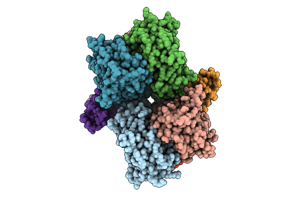
Organism: Homo sapiens
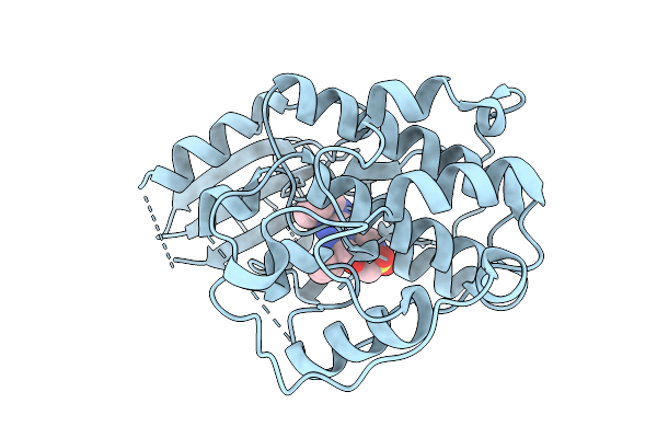
Method: X-RAY DIFFRACTION Release Date: 2025-09-10 Classification: TRANSFERASE Ligands: A1INI |
 |
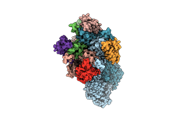
Organism: Homo sapiens
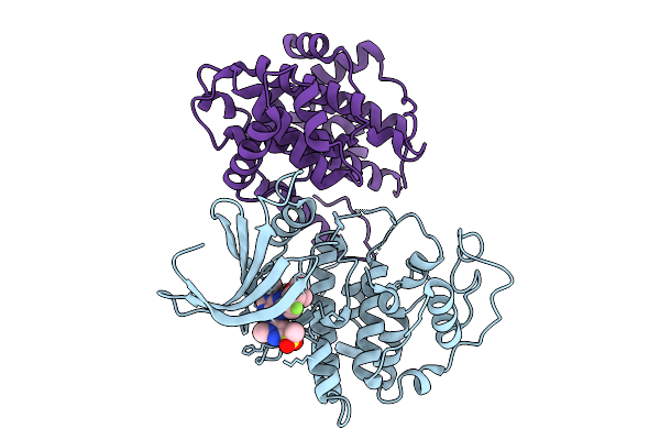
Method: X-RAY DIFFRACTION Release Date: 2025-09-10 Classification: TRANSFERASE Ligands: A1IOI |
 |
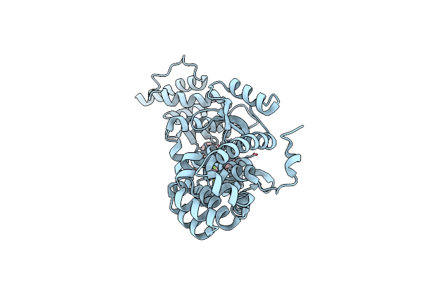
Organism: Homo sapiens
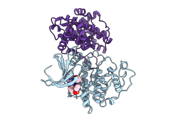
Method: X-RAY DIFFRACTION Release Date: 2025-09-10 Classification: TRANSFERASE Ligands: A1IOP |
 |
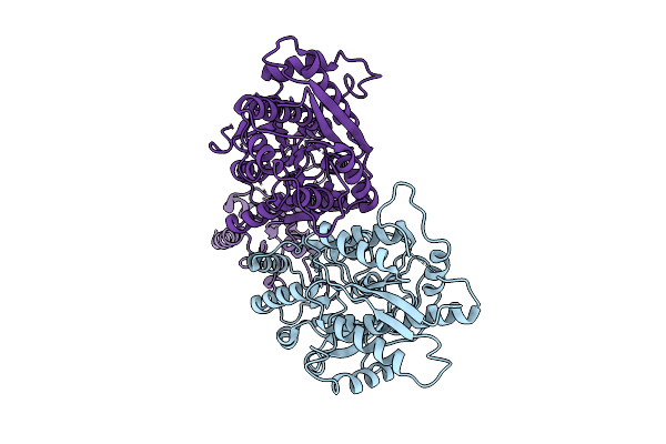
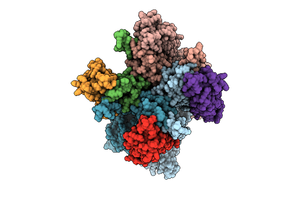
Crystal Structure Of (3R)-Hydroxyacyl-Acp Dehydratase Hadab Hetero-Dimer From Mycobacterium Tuberculosis Complexed With Substrate Palmitoyl-Coa
Organism: Mycobacterium tuberculosis h37rv
Method: X-RAY DIFFRACTION Release Date: 2025-07-30 Classification: LYASE Ligands: PLM, PEG, COA |
 |
The Ntd Dimer And The Interfacing Lbd Region Of The Ampar Complex Glua3(R439G,R163I)- Tarp Gamma2 In The Apo State.
Organism: Rattus norvegicus
Method: ELECTRON MICROSCOPY Release Date: 2025-07-30 Classification: MEMBRANE PROTEIN |
 |
The Tmd And The Lbd Region Of The Ampar Complex Glua3- Tarp Gamma2 In The Apo State.
Organism: Rattus norvegicus
Method: ELECTRON MICROSCOPY Release Date: 2025-07-09 Classification: MEMBRANE PROTEIN |
 |
The Ntd Dimer And The Interfacing Lbd Region Of The Ampar Complex Glua3- Tarp Gamma2 In The Open State.
Organism: Rattus norvegicus
Method: ELECTRON MICROSCOPY Release Date: 2025-07-09 Classification: MEMBRANE PROTEIN |
 |
The Ntd Dimer And The Interfacing Lbd Region Of The Ampar Complex Glua3- Tarp Gamma2 In The Apo State
Organism: Rattus norvegicus
Method: ELECTRON MICROSCOPY Release Date: 2025-07-09 Classification: MEMBRANE PROTEIN Ligands: NAG |
 |
The Ntd Dimer And The Interfacing Lbd Region Of The Ampar Complex Glua3- Tarp Gamma2 In The Desensitised State.
Organism: Rattus norvegicus
Method: ELECTRON MICROSCOPY Release Date: 2025-07-09 Classification: MEMBRANE PROTEIN Ligands: NAG |
 |
Organism: Rattus norvegicus
Method: ELECTRON MICROSCOPY Release Date: 2025-07-09 Classification: MEMBRANE PROTEIN |
 |
The Composite Map Of Of The Ampar Complex Glua3- Tarp Gamma2 In The Apo State.
Organism: Rattus norvegicus
Method: ELECTRON MICROSCOPY Release Date: 2025-07-09 Classification: MEMBRANE PROTEIN |
 |
Crystal Structure Of Transcriptional Regulator (Nrpr) From Streptococcus Salivarius K12
Organism: Streptococcus salivarius k12
Method: X-RAY DIFFRACTION Release Date: 2025-05-21 Classification: TRANSCRIPTION |
 |
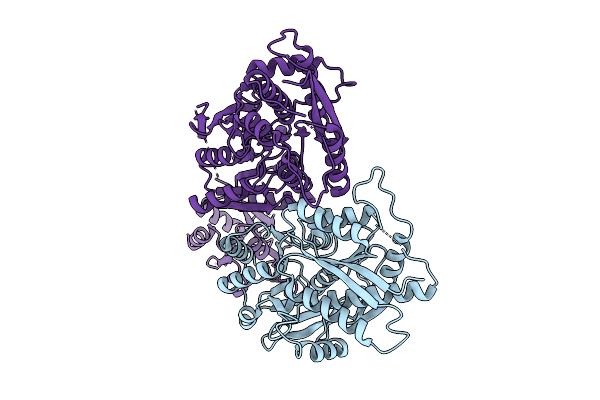
Crystal Structure Of Cbl-B In Complex With An Allosteric Inhibitor (Compound 31)
Organism: Homo sapiens
Method: X-RAY DIFFRACTION Resolution:1.87 Å Release Date: 2024-01-24 Classification: LIGASE Ligands: ZN, NA, WX9 |
 |
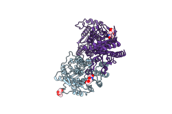
Crystal Structure Of Cbl-B In Complex With An Allosteric Inhibitor (Compound 9)
Organism: Homo sapiens
Method: X-RAY DIFFRACTION Resolution:1.42 Å Release Date: 2024-01-10 Classification: LIGASE Ligands: WUQ, ZN, NA |
 |
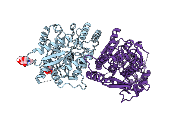
Crystal Structure Of Cbl-B In Complex With An Allosteric Inhibitor (Compound 8)
Organism: Homo sapiens
Method: X-RAY DIFFRACTION Resolution:2.20 Å Release Date: 2024-01-10 Classification: LIGASE Ligands: WX0, ZN, NA |
 |
Crystal Structure Of Cbl-B In Complex With An Allosteric Inhibitor (Compound 30)
Organism: Homo sapiens
Method: X-RAY DIFFRACTION Resolution:1.52 Å Release Date: 2024-01-10 Classification: LIGASE Ligands: ZN, NA, WUI |
 |
Crystal Structure Of An N Terminal Truncated Secreted Protein, Rv0398C From Mycobacterium Tuberculosis
Organism: Mycobacterium tuberculosis h37rv
Method: X-RAY DIFFRACTION Resolution:1.90 Å Release Date: 2023-06-28 Classification: IMMUNE SYSTEM Ligands: GOL |
 |
Crystal Structure Of Fada2 (Rv0243) From The Fatty Acid Metabolic Pathway Of Mycobacterium Tuberculosis
Organism: Mycobacterium tuberculosis h37rv
Method: X-RAY DIFFRACTION Resolution:2.90 Å Release Date: 2023-04-26 Classification: TRANSFERASE Ligands: SO4 |
 |
Organism: Vibrio harveyi
Method: X-RAY DIFFRACTION Resolution:2.00 Å Release Date: 2021-09-08 Classification: OXIDOREDUCTASE Ligands: PEG |
 |
Structure Of Dcn1 Bound To N-((4S,5S)-3-(Aminomethyl)-7-Ethyl-4-(4-Fluorophenyl)-6-Oxo-1-Phenyl-4,5,6,7-Tetrahydro-1H-Pyrazolo[3,4-B]Pyridin-5-Yl)-3-(Trifluoromethyl)Benzamide
Organism: Enterobacteria phage rb59, Homo sapiens
Method: X-RAY DIFFRACTION Resolution:1.57 Å Release Date: 2021-07-14 Classification: LIGASE/Inhibitor Ligands: YHP |