
Search Count: 24
All
Selected
 |
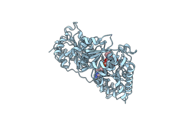
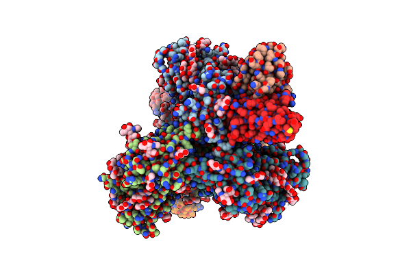
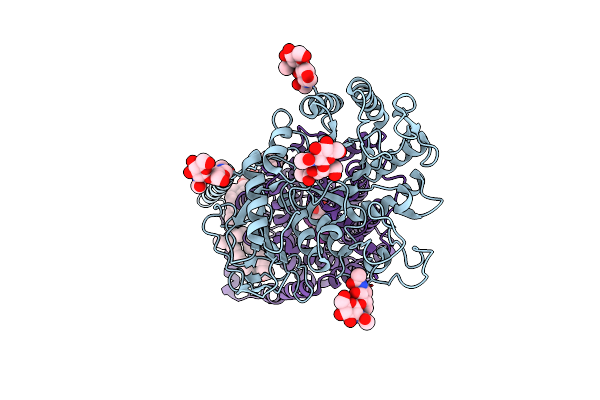
Corynebacterium Glutamicum Pyruvate:Quinone Oxidoreductase (Pqo) Purified From Bacteria Grown In Acetate Minimal Medium
Organism: Corynebacterium glutamicum atcc 13032
Method: X-RAY DIFFRACTION Resolution:1.22 Å Release Date: 2024-08-07 Classification: OXIDOREDUCTASE Ligands: FAD, TPP, MG |
 |
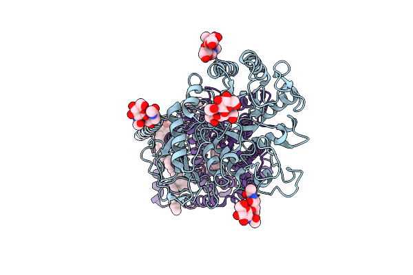
Pyruvate:Quinone Oxidoreductase (Pqo) From Corynebacterium Glutamicum Cs176
Organism: Corynebacterium glutamicum
Method: X-RAY DIFFRACTION Resolution:1.47 Å Release Date: 2024-08-07 Classification: OXIDOREDUCTASE Ligands: FAD |
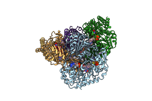 |
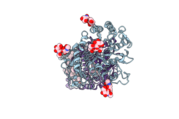
Corynebacterium Glutamicum Cs176 Pyruvate:Quinone Oxidoreductase (Pqo) In Complex With Fad And Thiamine Diphosphate-Magnesium Ion
Organism: Corynebacterium glutamicum
Method: X-RAY DIFFRACTION Resolution:1.86 Å Release Date: 2024-08-07 Classification: OXIDOREDUCTASE Ligands: TPP, MG, FAD |
 |
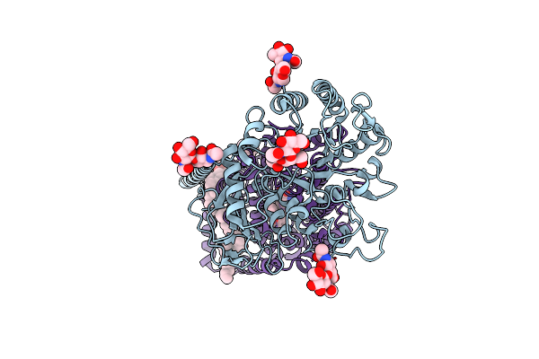
Corynebacterium Glutamicum Pyruvate:Quinone Oxidoreductase (Pqo), C-Terminal Truncated Construct
Organism: Corynebacterium glutamicum atcc 13032
Method: X-RAY DIFFRACTION Resolution:1.89 Å Release Date: 2024-08-07 Classification: OXIDOREDUCTASE Ligands: FAD, TPP, MG |
 |
Organism: Human immunodeficiency virus 1, Macaca mulatta
Method: ELECTRON MICROSCOPY Release Date: 2024-05-08 Classification: VIRAL PROTEIN/Immune System Ligands: NAG |
 |
Organism: Human immunodeficiency virus 1, Macaca mulatta
Method: ELECTRON MICROSCOPY Release Date: 2024-05-08 Classification: VIRAL PROTEIN/Immune System Ligands: NAG |
 |
Organism: Human immunodeficiency virus 1, Macaca mulatta
Method: ELECTRON MICROSCOPY Release Date: 2024-05-08 Classification: VIRAL PROTEIN/Immune System Ligands: NAG |
 |
Organism: Human immunodeficiency virus 1, Macaca mulatta
Method: ELECTRON MICROSCOPY Release Date: 2024-05-08 Classification: VIRAL PROTEIN/Immune System Ligands: NAG |
 |
Organism: Human immunodeficiency virus 1
Method: ELECTRON MICROSCOPY Release Date: 2024-05-08 Classification: VIRAL PROTEIN Ligands: NAG |
 |
Organism: Human immunodeficiency virus 1, Macaca mulatta
Method: ELECTRON MICROSCOPY Release Date: 2024-05-08 Classification: VIRAL PROTEIN/Immune System Ligands: NAG |
 |
Bg505 Sosip.V5.2 In Complex With The Monoclonal Antibody Rh4O9.8 (As Fab Fragment)
Organism: Human immunodeficiency virus 1, Macaca mulatta
Method: ELECTRON MICROSCOPY Release Date: 2022-01-26 Classification: VIRAL PROTEIN Ligands: NAG |
 |
Bg505 Sosip Md39 In Complex With The Monoclonal Antibodies Rh.33104 Mab.1 And Rm20A3
Organism: Macaca mulatta, Human immunodeficiency virus 1
Method: ELECTRON MICROSCOPY Release Date: 2022-01-26 Classification: VIRAL PROTEIN Ligands: NAG |
 |
Bg505 Sosip.V5.2(7S) In Complex With The Monoclonal Antibodies Rh.33172 Mab.1 And Rm19R
Organism: Macaca mulatta, Human immunodeficiency virus
Method: ELECTRON MICROSCOPY Release Date: 2022-01-26 Classification: VIRAL PROTEIN Ligands: NAG |
 |
Organism: Homo sapiens
Method: ELECTRON MICROSCOPY Release Date: 2021-03-10 Classification: MEMBRANE PROTEIN Ligands: 3PH, HNX, Y01 |
 |
Organism: Homo sapiens
Method: ELECTRON MICROSCOPY Release Date: 2021-03-10 Classification: MEMBRANE PROTEIN Ligands: 3PH, HO0, Y01 |
 |
Organism: Homo sapiens
Method: ELECTRON MICROSCOPY Release Date: 2021-03-10 Classification: MEMBRANE PROTEIN Ligands: 3PH, HO9, Y01 |
 |
Organism: Homo sapiens
Method: ELECTRON MICROSCOPY Release Date: 2021-03-10 Classification: MEMBRANE PROTEIN Ligands: TYI, Y01, 3PH |
 |
Crystal Structure Of Ioma-Class Clk31 Fab From An Hiv-1 Naive Donor In Complex With A Germline-Targeting Gp120 Engineered Outer Domain Eod-Gt8 At 2.6 A
Organism: Homo sapiens
Method: X-RAY DIFFRACTION Resolution:2.60 Å Release Date: 2019-01-16 Classification: IMMUNE SYSTEM Ligands: GOL |
 |
Organism: Enterobacter lignolyticus
Method: X-RAY DIFFRACTION Resolution:2.16 Å Release Date: 2018-06-27 Classification: TRANSCRIPTION/DNA Ligands: HEZ |
 |
Organism: Enterobacter lignolyticus
Method: X-RAY DIFFRACTION Resolution:2.93 Å Release Date: 2018-06-27 Classification: TRANSCRIPTION Ligands: MGR |