
Search Count: 47
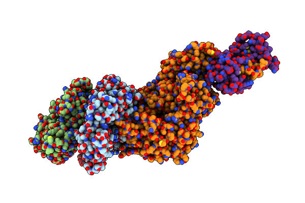 |
Organism: Mus musculus, Oryctolagus cuniculus
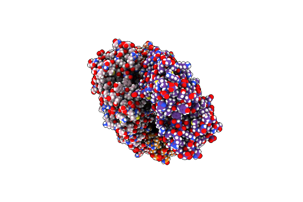
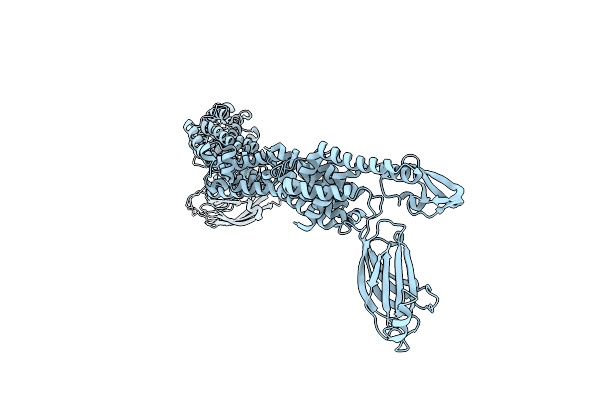
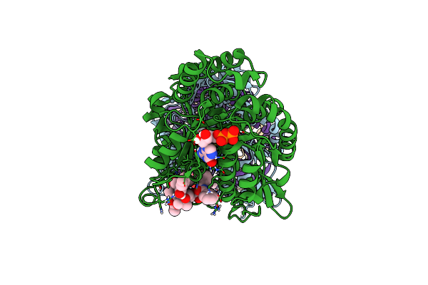
Method: ELECTRON MICROSCOPY Release Date: 2025-02-12 Classification: MOTOR PROTEIN Ligands: ADP, MG |
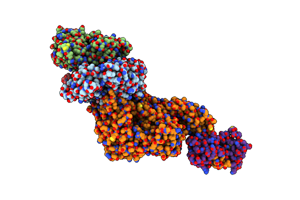 |
Organism: Mus musculus, Oryctolagus cuniculus
Method: ELECTRON MICROSCOPY Release Date: 2025-02-12 Classification: MOTOR PROTEIN Ligands: ADP, MG |
 |
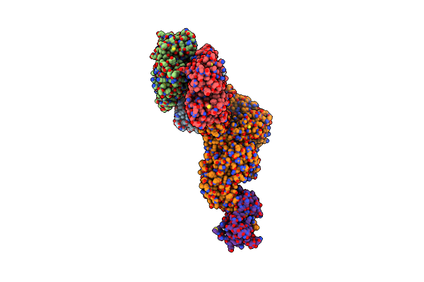
Organism: Mus musculus, Oryctolagus cuniculus
Method: ELECTRON MICROSCOPY Release Date: 2025-02-12 Classification: MOTOR PROTEIN Ligands: ADP, MG |
 |
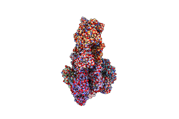
Organism: Mus musculus, Oryctolagus cuniculus
Method: ELECTRON MICROSCOPY Release Date: 2025-02-12 Classification: MOTOR PROTEIN Ligands: ADP, MG |
 |
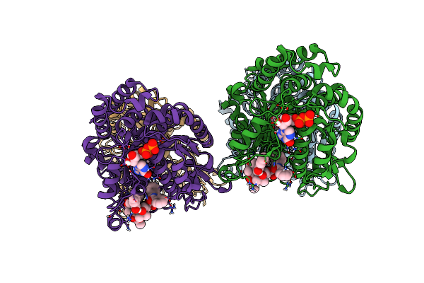
Organism: Schizosaccharomyces pombe, Oryctolagus cuniculus
Method: ELECTRON MICROSCOPY Release Date: 2024-01-31 Classification: CYTOSOLIC PROTEIN Ligands: ADP, MG, ATP |
 |
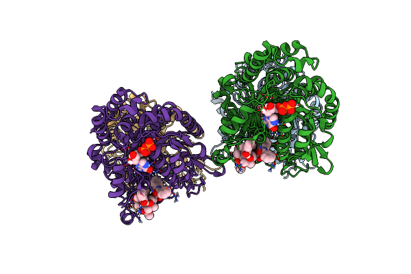
Organism: Schizosaccharomyces pombe, Oryctolagus cuniculus
Method: ELECTRON MICROSCOPY Release Date: 2024-01-31 Classification: CYTOSOLIC PROTEIN Ligands: ADP, MG, BEF, ATP |
 |
Organism: Oryctolagus cuniculus
Method: ELECTRON MICROSCOPY Release Date: 2024-01-31 Classification: CYTOSOLIC PROTEIN Ligands: ADP, MG |
 |
Organism: Oryctolagus cuniculus
Method: ELECTRON MICROSCOPY Release Date: 2024-01-31 Classification: CYTOSOLIC PROTEIN Ligands: ADP, MG, BEF |
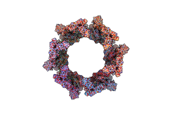 |
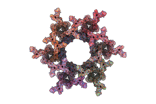
The Hexagonal Organization Of Munc13-1 C1-C2B-Mun-C2C Domains Between Lipid Bilayers
Organism: Rattus norvegicus
Method: ELECTRON MICROSCOPY Release Date: 2022-02-09 Classification: EXOCYTOSIS |
 |
Organism: Mus musculus
Method: ELECTRON MICROSCOPY Release Date: 2022-02-09 Classification: EXOCYTOSIS |
 |
Organism: Rattus norvegicus
Method: ELECTRON MICROSCOPY Release Date: 2022-02-09 Classification: EXOCYTOSIS |
 |
Organism: Rattus norvegicus
Method: ELECTRON MICROSCOPY Release Date: 2022-02-09 Classification: EXOCYTOSIS |
 |
Organism: Rattus norvegicus
Method: ELECTRON MICROSCOPY Release Date: 2022-02-09 Classification: EXOCYTOSIS |
 |
Low Curvature Lateral Interaction Within A 13-Protofilament, Taxol Stabilized Microtubule
Organism: Bos taurus
Method: ELECTRON MICROSCOPY Release Date: 2020-05-20 Classification: STRUCTURAL PROTEIN Ligands: GTP, MG, GDP, TA1 |
 |
High Curvature Lateral Interaction Within A 13-Protofilament, Taxol Stabilized Microtubule
Organism: Bos taurus
Method: ELECTRON MICROSCOPY Release Date: 2020-05-20 Classification: STRUCTURAL PROTEIN Ligands: GTP, MG, GDP, TA1 |
 |
Organism: Bos taurus
Method: ELECTRON MICROSCOPY Release Date: 2020-05-20 Classification: STRUCTURAL PROTEIN Ligands: GTP, MG, GDP, TA1 |
 |
Monomeric Kinesin-1 Motor Domain In No-Nucleotide State Bound To Gmpcpp-Stabilized Microtubule
Organism: Homo sapiens, Bos taurus
Method: ELECTRON MICROSCOPY Release Date: 2020-04-15 Classification: MOTOR PROTEIN Ligands: G2P, MG, GTP |
 |
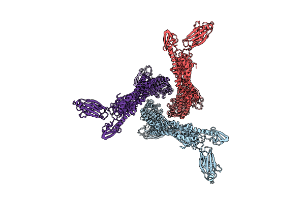
Rigid Body Fitting Of Flagellin Flab, And Flagellar Coiling Proteins, Fcpa And Fcpb, Into A 10 Angstrom Structure Of The Asymmetric Flagellar Filament Purified From Leptospira Biflexa Patoc Wt Cells Resolved Via Subtomogram Averaging
Organism: Leptospira biflexa serovar patoc (strain patoc 1 / atcc 23582 / paris)
Method: ELECTRON MICROSCOPY Release Date: 2020-03-25 Classification: STRUCTURAL PROTEIN |
 |
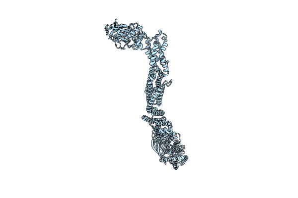
Cryo-Em Structure Of Microtubule-Bound Klp61F Motor Domain In The Amppnp State
Organism: Drosophila melanogaster, Sus scrofa
Method: ELECTRON MICROSCOPY Release Date: 2020-02-19 Classification: MOTOR PROTEIN Ligands: GTP, MG, GDP, ANP |
 |
Cryo-Em Structure Of Microtubule-Bound Klp61F Motor With Tail Domain In The Nucleotide-Free State
Organism: Drosophila melanogaster, Sus scrofa
Method: ELECTRON MICROSCOPY Release Date: 2020-02-19 Classification: MOTOR PROTEIN Ligands: GTP, MG, GDP |