
Search Count: 19
All
Selected
 |
Cryo-Em Structure Of A Foamy Virus Fusion Glycoprotein Stabilized In The Prefusion Conformation
Organism: Simian foamy virus
Method: ELECTRON MICROSCOPY Release Date: 2024-10-23 Classification: VIRUS Ligands: NAG |
 |
Cryo-Em Structure Of A Foamy Virus Fusion Glycoprotein In The Postfusion Conformation
Organism: Simian foamy virus
Method: ELECTRON MICROSCOPY Release Date: 2024-10-23 Classification: VIRUS Ligands: NAG |
 |
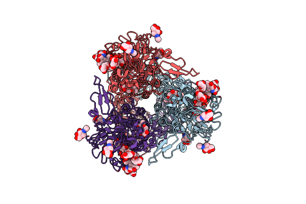
Organism: Eastern chimpanzee simian foamy virus
Method: ELECTRON MICROSCOPY Resolution:2.91 Å Release Date: 2024-07-10 Classification: VIRAL PROTEIN Ligands: NAG, CLR, PTY |
 |
Organism: Eastern chimpanzee simian foamy virus
Method: ELECTRON MICROSCOPY Resolution:3.70 Å Release Date: 2024-07-10 Classification: VIRAL PROTEIN Ligands: CLR, PTY, NAG |
 |
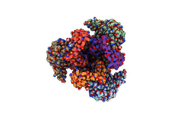
In Situ Cryoem Structure Of The Prototype Foamy Virus Capsid, Icosahedral Map
Organism: Eastern chimpanzee simian foamy virus
Method: ELECTRON MICROSCOPY Resolution:3.89 Å Release Date: 2024-07-10 Classification: VIRAL PROTEIN |
 |
In Situ Cryoem Structure Of The Prototype Foamy Virus Capsid, Pentamer Localised Reconstruction
Organism: Eastern chimpanzee simian foamy virus
Method: ELECTRON MICROSCOPY Resolution:3.40 Å Release Date: 2024-07-10 Classification: VIRAL PROTEIN |
 |
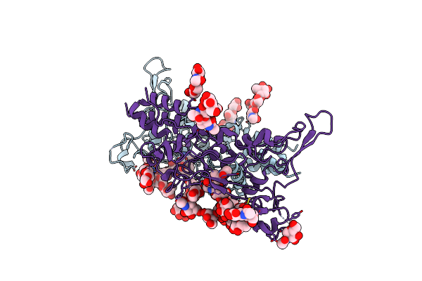
In Situ Cryoem Structure Of The Prototype Foamy Virus Capsid, Hexamer 1 Localised Reconstruction
Organism: Eastern chimpanzee simian foamy virus
Method: ELECTRON MICROSCOPY Resolution:3.40 Å Release Date: 2024-07-10 Classification: VIRAL PROTEIN |
 |
In Situ Cryoem Structure Of The Prototype Foamy Virus Capsid, Hexamer 2 Localised Reconstruction
Organism: Eastern chimpanzee simian foamy virus
Method: ELECTRON MICROSCOPY Resolution:3.50 Å Release Date: 2024-07-10 Classification: VIRAL PROTEIN |
 |
In Situ Subtomogram Average Of Prototype Foamy Virus Env Pentamer Of Trimers
Organism: Eastern chimpanzee simian foamy virus
Method: ELECTRON MICROSCOPY Resolution:11.90 Å Release Date: 2024-07-10 Classification: VIRAL PROTEIN Ligands: CLR, NAG, PTY |
 |
In Situ Subtomogram Average Of Prototype Foamy Virus Env Hexamer Of Trimers
Organism: Eastern chimpanzee simian foamy virus
Method: ELECTRON MICROSCOPY Resolution:10.00 Å Release Date: 2024-07-10 Classification: VIRAL PROTEIN Ligands: NAG, CLR, PTY |
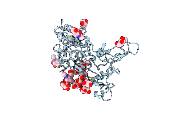 |
X-Ray Structure Of The Deglycosylated Receptor Binding Domain Of Env Glycoprotein Of Simian Foamy Virus
Organism: Simian foamy virus
Method: X-RAY DIFFRACTION Resolution:2.57 Å Release Date: 2023-02-22 Classification: VIRAL PROTEIN Ligands: NAG |
 |
X-Ray Structure Of The Receptor Binding Domain Of Env Glycoprotein Of Simian Foamy Virus
Organism: Simian foamy virus
Method: X-RAY DIFFRACTION Resolution:2.80 Å Release Date: 2023-02-22 Classification: VIRAL PROTEIN Ligands: NAG |
 |
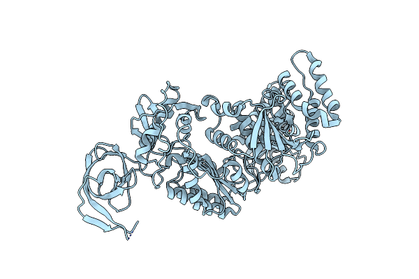
Crystal Structure Of Prototype Foamy Virus Protease-Reverse Transcriptase Csh Mutant (Selenomethionine-Labeled)
Organism: Eastern chimpanzee simian foamy virus
Method: X-RAY DIFFRACTION Resolution:3.00 Å Release Date: 2021-08-11 Classification: VIRAL PROTEIN Ligands: CA |
 |
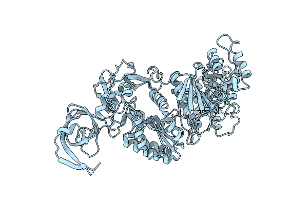
Crystal Structure Of Prototype Foamy Virus Protease-Reverse Transcriptase (Native)
Organism: Eastern chimpanzee simian foamy virus
Method: X-RAY DIFFRACTION Resolution:2.90 Å Release Date: 2021-08-11 Classification: VIRAL PROTEIN Ligands: CA |
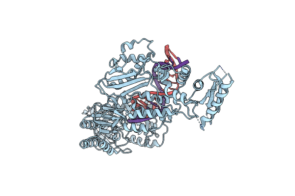 |
Structure Of The Foamy Viral Protease-Reverse Transcriptase In Complex With Rna/Dna Hybrid.
Organism: White-tufted-ear marmoset simian foamy virus, Synthetic construct
Method: X-RAY DIFFRACTION Resolution:3.10 Å Release Date: 2021-06-30 Classification: VIRAL PROTEIN Ligands: HOH |
 |
Structure Of The Foamy Viral Protease-Reverse Transcriptase Drh In Complex With Ds Dna.
Organism: White-tufted-ear marmoset simian foamy virus, Synthetic construct
Method: X-RAY DIFFRACTION Resolution:3.09 Å Release Date: 2021-06-30 Classification: VIRAL PROTEIN |
 |
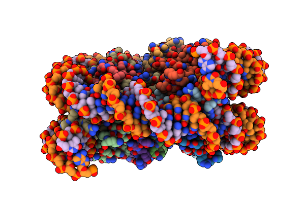
Structure Of The Foamy Viral Protease-Reverse Transcriptase In Complex With Dsdna.
Organism: White-tufted-ear marmoset simian foamy virus, Synthetic construct
Method: ELECTRON MICROSCOPY Release Date: 2021-06-30 Classification: VIRAL PROTEIN |
 |
Organism: Xenopus laevis, Simian foamy virus, Escherichia coli
Method: X-RAY DIFFRACTION Resolution:2.80 Å Release Date: 2017-05-10 Classification: DNA BINDING PROTEIN Ligands: MN |
 |
Organism: Simian foamy virus type 1
Method: SOLUTION NMR Release Date: 2008-06-03 Classification: HYDROLASE |