
Search Count: 38
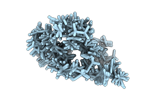 |
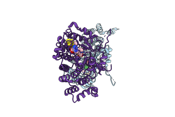
Staphylococcus Aureus Moaa C-Terminal Tail Peptide In Complex With G340A-Moaa
Organism: Staphylococcus aureus
Method: SOLUTION NMR Release Date: 2025-11-26 Classification: BIOSYNTHETIC PROTEIN |
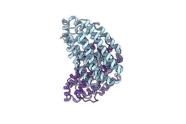 |
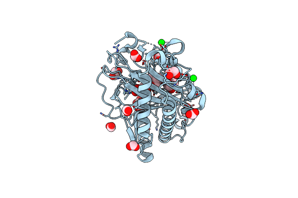
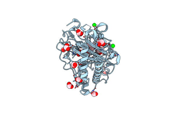
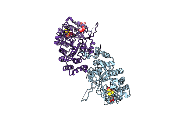
X-Ray Crystal Structure Of Francisella Hispaniensis Apo Ribonucleotide Reductase Beta Subunit
Organism: Francisella hispaniensis
Method: X-RAY DIFFRACTION Release Date: 2025-09-17 Classification: OXIDOREDUCTASE Ligands: CL |
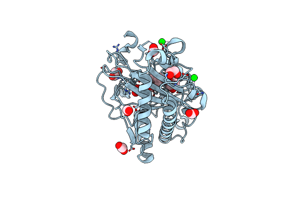 |
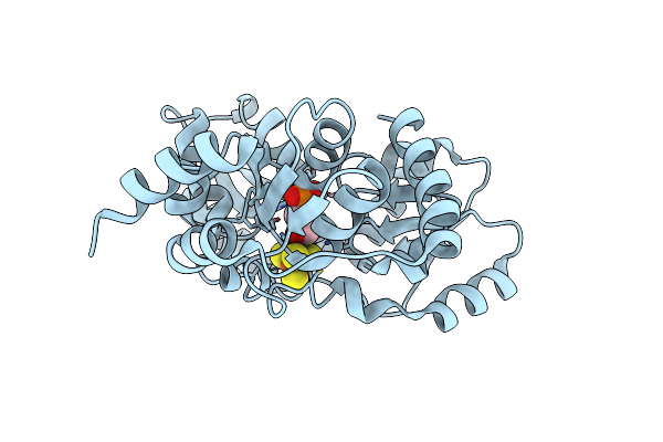
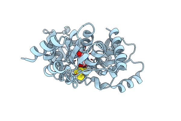
Structure Of Hyoscyamine 6-Beta Hydroxylase In Complex With Iron, 2-Oxoglutarate, And Hyoscyamine
Organism: Atropa belladonna
Method: X-RAY DIFFRACTION Resolution:1.53 Å Release Date: 2023-11-22 Classification: OXIDOREDUCTASE Ligands: SR, EDO, FMT, FE2, AKG, HYO |
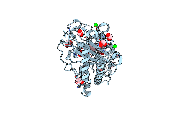 |
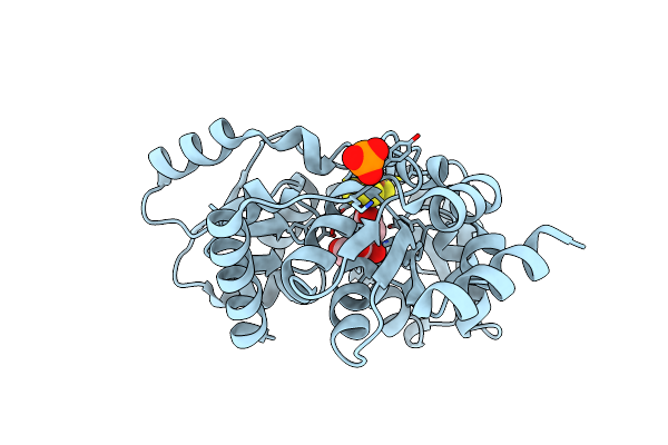
Structure Of Hyoscyamine 6-Beta Hydroxylase In Complex With Vanadyl, Succinate, And Hyoscyamine
Organism: Atropa belladonna
Method: X-RAY DIFFRACTION Resolution:1.79 Å Release Date: 2023-11-22 Classification: OXIDOREDUCTASE Ligands: EDO, HYO, SIN, FMT, V, O, SR |
 |
Structure Of Hyoscyamine 6-Beta Hydroxylase In Complex With Iron, Succinate, And 6-Oh-Hyoscyamine
Organism: Atropa belladonna
Method: X-RAY DIFFRACTION Resolution:1.58 Å Release Date: 2023-11-22 Classification: OXIDOREDUCTASE Ligands: FE2, EDO, FMT, OVR, SIN, SR |
 |
Structure Of Hyoscyamine 6-Beta Hydroxylase In Complex With Iron, 2-Oxoglutarate, And 6-Oh-Hyoscyamine
Organism: Atropa belladonna
Method: X-RAY DIFFRACTION Resolution:1.53 Å Release Date: 2023-11-22 Classification: OXIDOREDUCTASE Ligands: FE2, EDO, FMT, AKG, OVR, SR |
 |
Structure Of Hyoscyamine 6-Beta Hydroxylase In Complex With Vanadyl, Succinate, And 6-Oh-Hyoscyamine
Organism: Atropa belladonna
Method: X-RAY DIFFRACTION Resolution:1.79 Å Release Date: 2023-11-22 Classification: OXIDOREDUCTASE Ligands: EDO, SIN, FMT, OVR, V, O, SR |
 |
Structure Of Hyoscyamine 6-Beta Hydroxylase In Complex With Iron, Succinate, And Scopolamine
Organism: Atropa belladonna
Method: X-RAY DIFFRACTION Resolution:1.72 Å Release Date: 2023-11-22 Classification: OXIDOREDUCTASE Ligands: FE2, EDO, FMT, OW0, SIN, SR |
 |
Structure Of L289F Hyoscyamine 6-Beta Hydroxylase In Complex With Iron, 2-Oxoglutarate, And Hyoscyamine
Organism: Atropa belladonna
Method: X-RAY DIFFRACTION Resolution:1.53 Å Release Date: 2023-11-22 Classification: OXIDOREDUCTASE Ligands: FE2, EDO, FMT, HYO, AKG, SR |
 |
Structure Of L289F Hyoscyamine 6-Beta Hydroxylase In Complex With Vanadyl, Succinate, And Hyoscyamine
Organism: Atropa belladonna
Method: X-RAY DIFFRACTION Resolution:1.53 Å Release Date: 2023-11-22 Classification: OXIDOREDUCTASE Ligands: EDO, FMT, HYO, SIN, V, O, SR |
 |
Structure Of L289F Hyoscyamine 6-Beta Hydroxylase In Complex With Iron, 2-Oxoglutarate, And 6-Oh-Hyoscyamine
Organism: Atropa belladonna
Method: X-RAY DIFFRACTION Resolution:1.56 Å Release Date: 2023-11-22 Classification: OXIDOREDUCTASE Ligands: FE2, EDO, FMT, AKG, OVR, SR |
 |
Structure Of L289F Hyoscyamine 6-Beta Hydroxylase In Complex With Vanadyl, Succinate, And 6-Oh-Hyoscyamine
Organism: Atropa belladonna
Method: X-RAY DIFFRACTION Resolution:2.03 Å Release Date: 2023-11-22 Classification: OXIDOREDUCTASE Ligands: EDO, FMT, SIN, OVR, V, SR |
 |
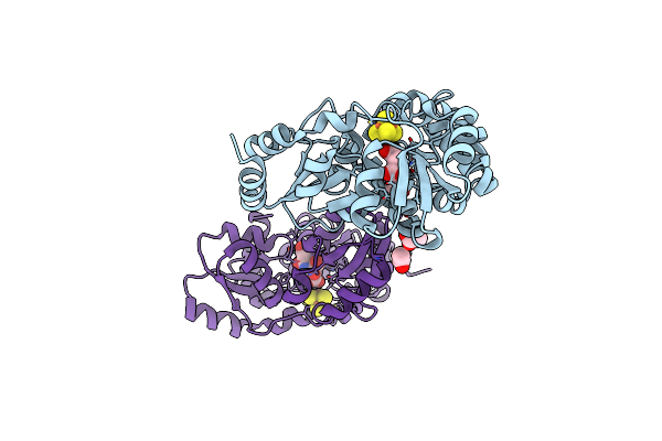
X-Ray Crystal Structure Of Mqne From Pedobacter Heparinus In Complex With Aminofutalosine And Methionine
Organism: Pedobacter heparinus (strain atcc 13125 / dsm 2366 / cip 104194 / jcm 7457 / nbrc 12017 / ncimb 9290 / nrrl b-14731 / him 762-3)
Method: X-RAY DIFFRACTION Resolution:2.14 Å Release Date: 2020-07-15 Classification: BIOSYNTHETIC PROTEIN Ligands: SF4, MET, V47, CL |
 |
Organism: Pedobacter heparinus (strain atcc 13125 / dsm 2366 / cip 104194 / jcm 7457 / nbrc 12017 / ncimb 9290 / nrrl b-14731 / him 762-3)
Method: X-RAY DIFFRACTION Resolution:1.59 Å Release Date: 2020-07-15 Classification: BIOSYNTHETIC PROTEIN Ligands: SF4, TAR |
 |
An Unexpected Intermediate In The Reaction Catalyzed By Quinolinate Synthase
Organism: Pyrococcus horikoshii (strain atcc 700860 / dsm 12428 / jcm 9974 / nbrc 100139 / ot-3)
Method: X-RAY DIFFRACTION Resolution:1.60 Å Release Date: 2019-10-16 Classification: TRANSFERASE Ligands: 13P, SF4 |
 |
An Unexpected Intermediate In The Reaction Catalyzed By Quinolinate Synthase
Organism: Pyrococcus horikoshii (strain atcc 700860 / dsm 12428 / jcm 9974 / nbrc 100139 / ot-3)
Method: X-RAY DIFFRACTION Resolution:1.65 Å Release Date: 2019-10-16 Classification: TRANSFERASE Ligands: SF4, 5XR, PO4 |
 |
Crystallographic Capture Of Quinolinate Synthase (Nada) From Pyrococcus Horikoshii In Its Substrates And Product-Bound States
Organism: Pyrococcus horikoshii (strain atcc 700860 / dsm 12428 / jcm 9974 / nbrc 100139 / ot-3)
Method: X-RAY DIFFRACTION Resolution:2.15 Å Release Date: 2019-08-21 Classification: TRANSFERASE Ligands: SF4, DYA |
 |
An Unexpected Intermediate In The Reaction Catalyzed By Quinolinate Synthase
Organism: Pyrococcus horikoshii (strain atcc 700860 / dsm 12428 / jcm 9974 / nbrc 100139 / ot-3)
Method: X-RAY DIFFRACTION Resolution:2.20 Å Release Date: 2019-08-21 Classification: TRANSFERASE Ligands: SF4, ACT, 5XW, CL, 5XR |
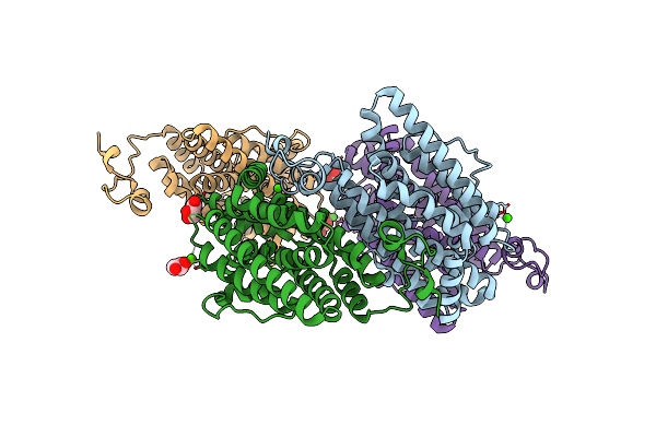 |
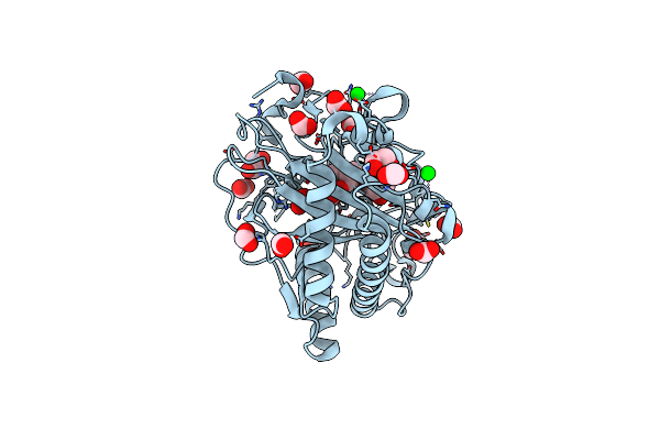
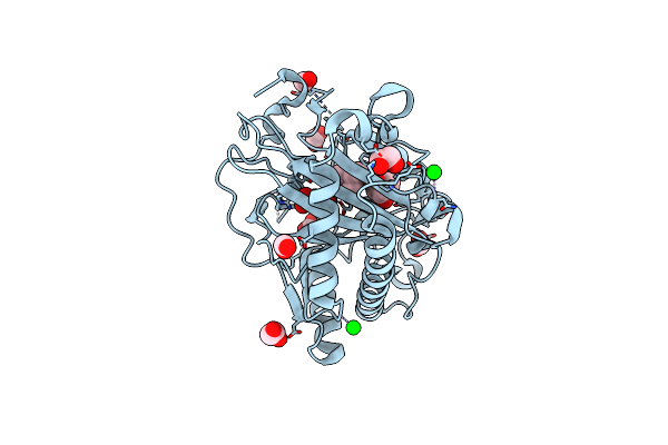
Crystal Structure Of The Class Ie Ribonucleotide Reductase Beta Subunit From Aerococcus Urinae In Unactivated Form
Organism: Aerococcus urinae (strain acs-120-v-col10a)
Method: X-RAY DIFFRACTION Resolution:1.58 Å Release Date: 2018-09-19 Classification: OXIDOREDUCTASE Ligands: CA, GOL |
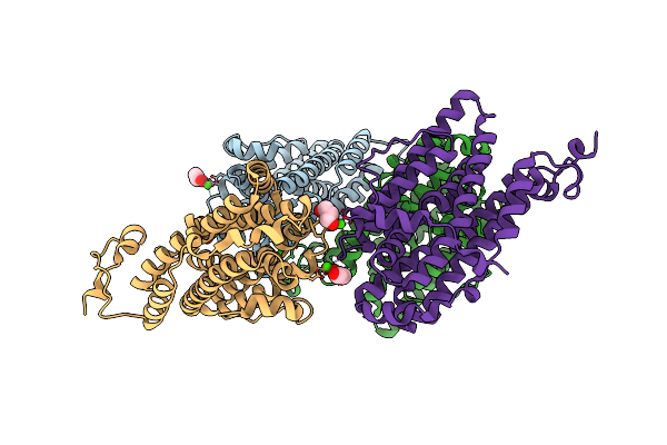 |
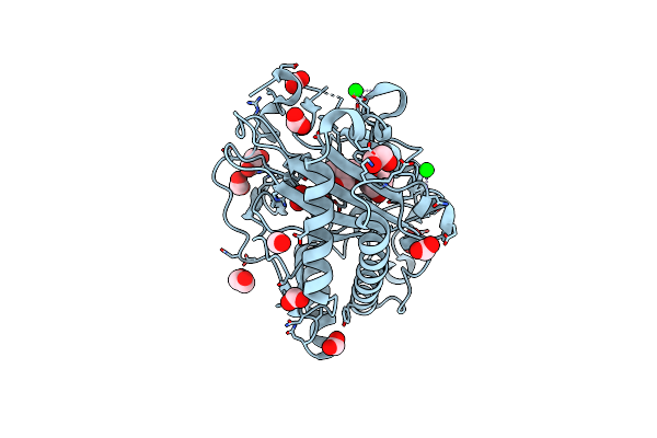
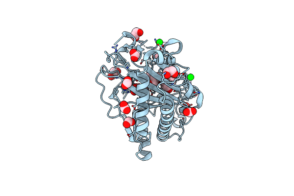
Crystal Structure Of The Class Ie Ribonucleotide Reductase Beta Subunit From Aerococcus Urinae In Activated Form
Organism: Aerococcus urinae (strain acs-120-v-col10a)
Method: X-RAY DIFFRACTION Resolution:1.59 Å Release Date: 2018-09-19 Classification: OXIDOREDUCTASE Ligands: CA, GOL |