Search Count: 45
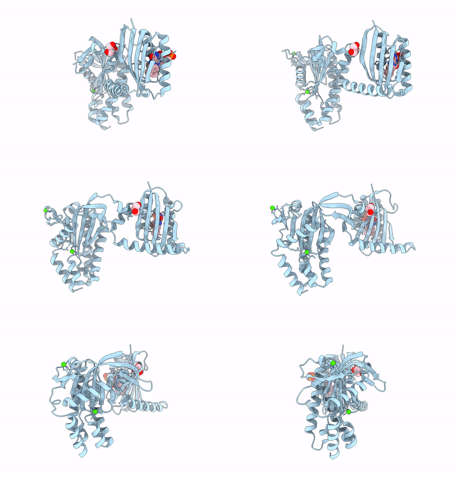 |
Organism: Oscillatoria acuminata
Method: X-RAY DIFFRACTION Resolution:1.95 Å Release Date: 2025-02-26 Classification: LYASE Ligands: FMN, GOL, CA |
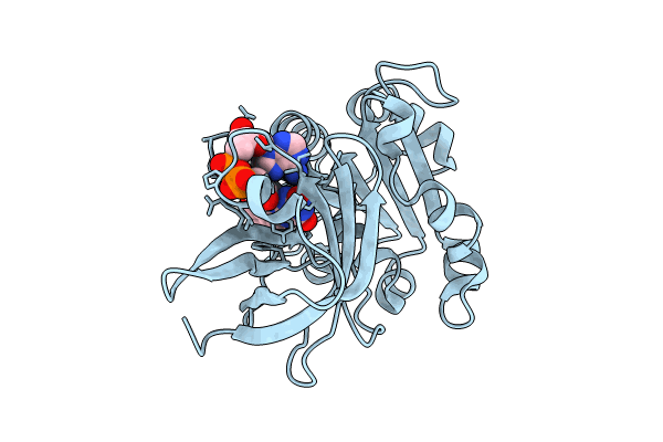 |
Room Temperature Structure Of Fad-Containing Ferrodoxin-Nadp Reductase From Brucella Ovis At Euxfel
Organism: Brucella ovis atcc 25840
Method: X-RAY DIFFRACTION Resolution:1.90 Å Release Date: 2024-11-27 Classification: OXIDOREDUCTASE Ligands: FAD |
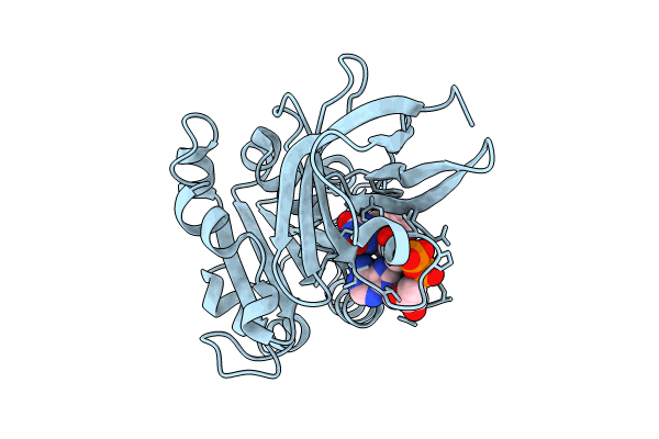 |
Room Temperature Structure Of Fad-Containing Ferrodoxin-Nadp Reductase From Brucella Ovis At Lcls
Organism: Brucella ovis atcc 25840
Method: X-RAY DIFFRACTION Resolution:2.20 Å Release Date: 2024-11-27 Classification: OXIDOREDUCTASE Ligands: FAD |
 |
Structure Of The Lysinibacillus Sphaericus Tpp49Aa1 Pesticidal Protein At Ph 3
Organism: Lysinibacillus sphaericus
Method: X-RAY DIFFRACTION Resolution:1.78 Å Release Date: 2023-11-01 Classification: TOXIN |
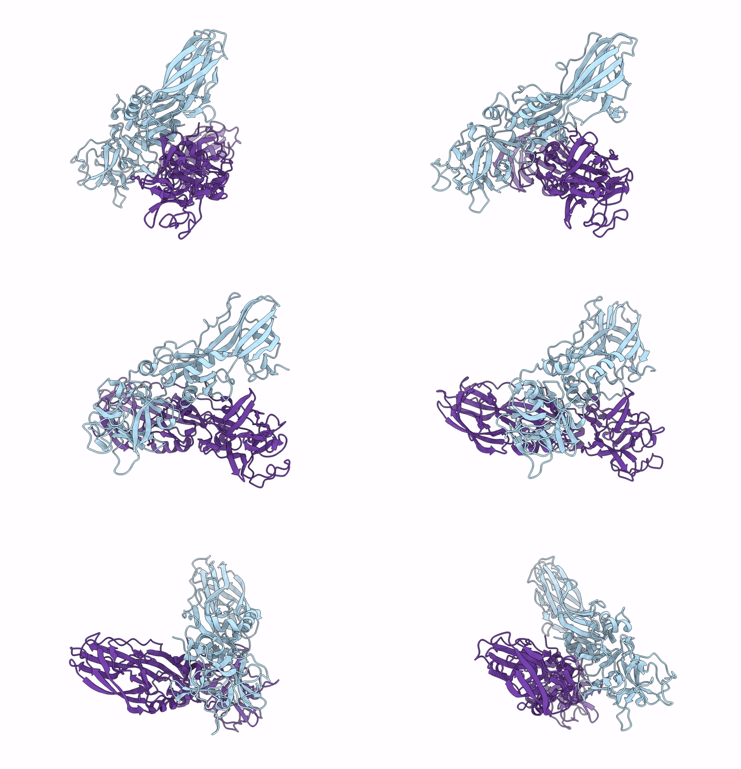 |
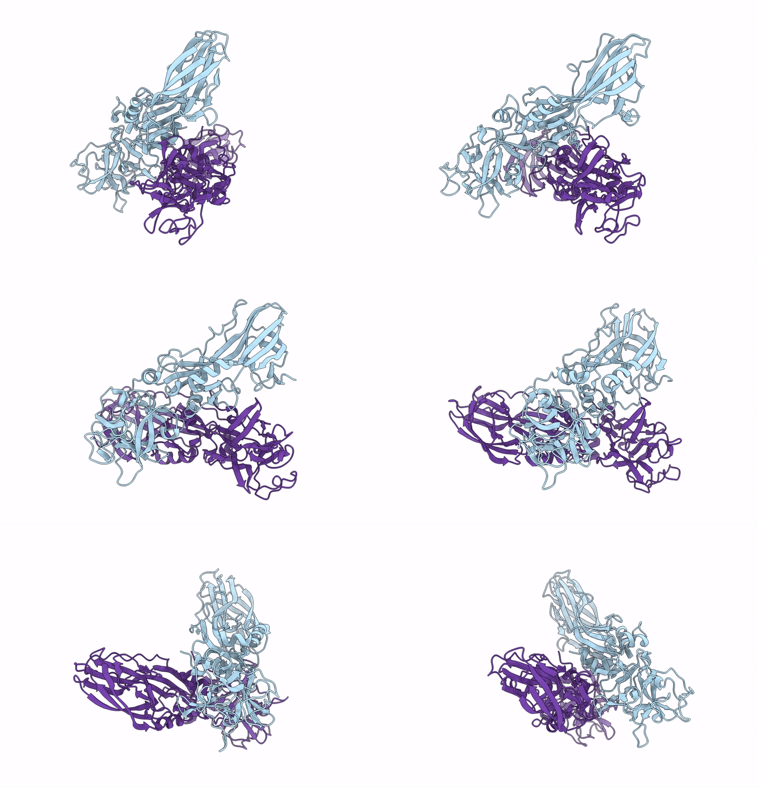
Structure Of The Lysinibacillus Sphaericus Tpp49Aa1 Pesticidal Protein At Ph 7
Organism: Lysinibacillus sphaericus
Method: X-RAY DIFFRACTION Resolution:1.62 Å Release Date: 2023-11-01 Classification: TOXIN |
 |
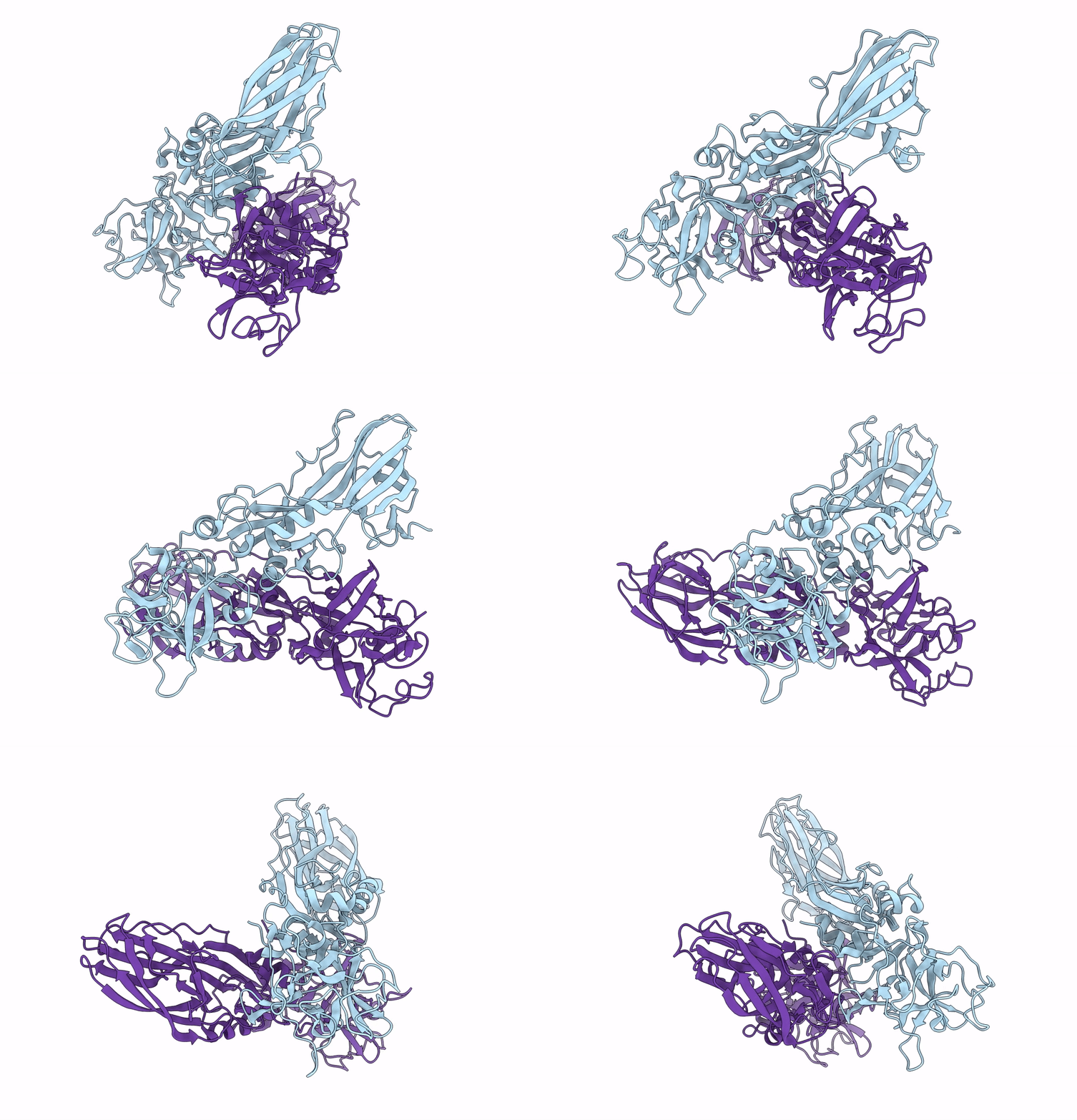
Structure Of The Lysinibacillus Sphaericus Tpp49Aa1 Pesticidal Protein At Ph 11
Organism: Lysinibacillus sphaericus
Method: X-RAY DIFFRACTION Resolution:1.75 Å Release Date: 2023-11-01 Classification: TOXIN |
 |
The Structure Of Natural Crystals Of The Lysinibacillus Sphaericus Tpp49Aa1 Pesticidal Protein Elucidated Using Serial Femtosecond Crystallography At An X-Ray Free Electron Laser
Organism: Lysinibacillus sphaericus
Method: X-RAY DIFFRACTION Resolution:2.20 Å Release Date: 2023-05-17 Classification: TOXIN |
 |
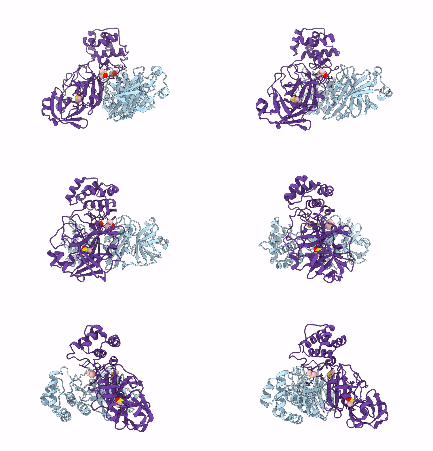
Crystal Structure Of Sars-Cov-2 Main Protease In Orthorhombic Space Group P212121
Organism: Severe acute respiratory syndrome coronavirus 2
Method: X-RAY DIFFRACTION Resolution:1.65 Å Release Date: 2023-03-22 Classification: HYDROLASE Ligands: CL, DMS, MLI |
 |
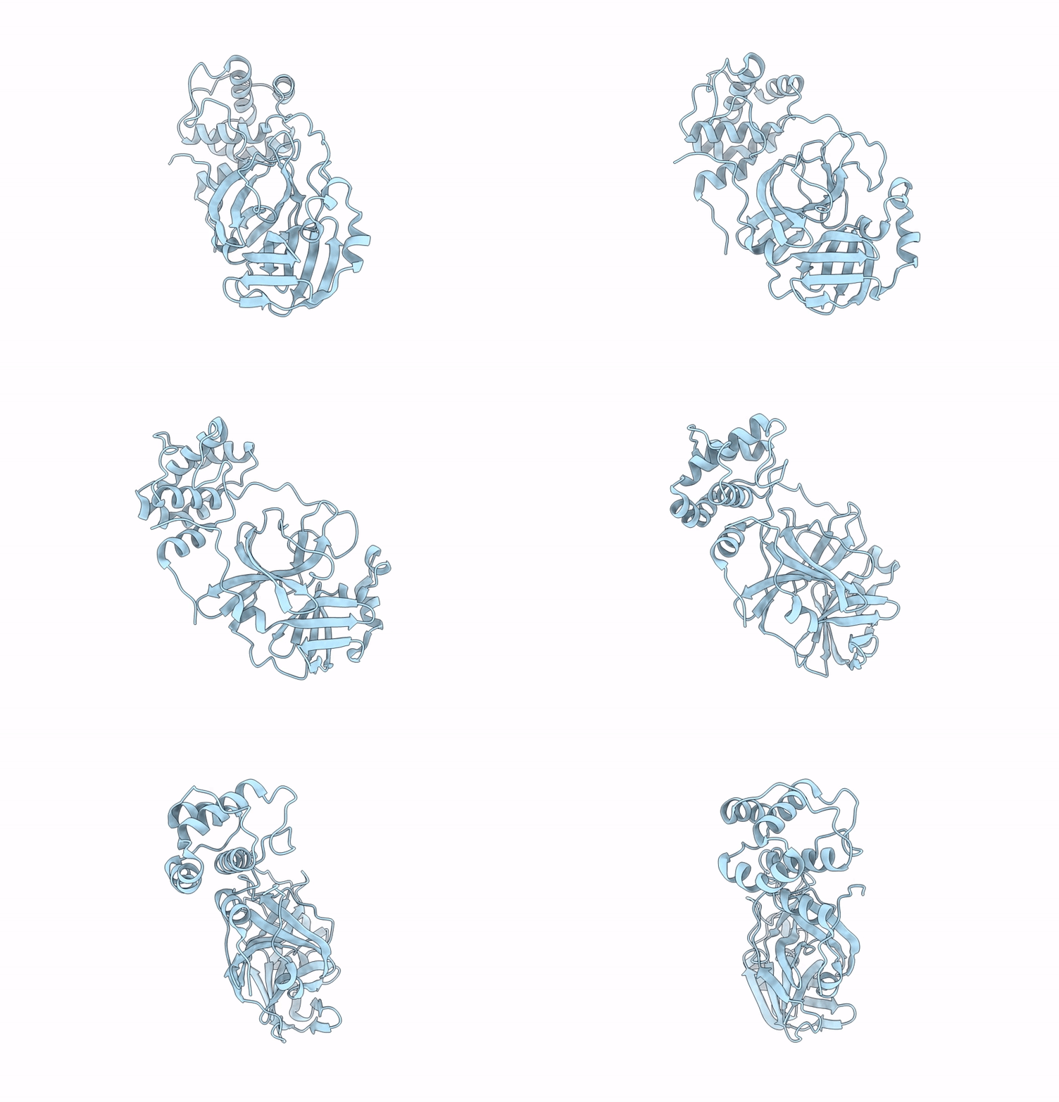
Organism: Severe acute respiratory syndrome coronavirus 2
Method: X-RAY DIFFRACTION Resolution:2.25 Å Release Date: 2023-01-25 Classification: VIRAL PROTEIN Ligands: DMS |
 |
Organism: Severe acute respiratory syndrome coronavirus 2
Method: X-RAY DIFFRACTION Resolution:1.75 Å Release Date: 2023-01-18 Classification: VIRAL PROTEIN Ligands: CL |
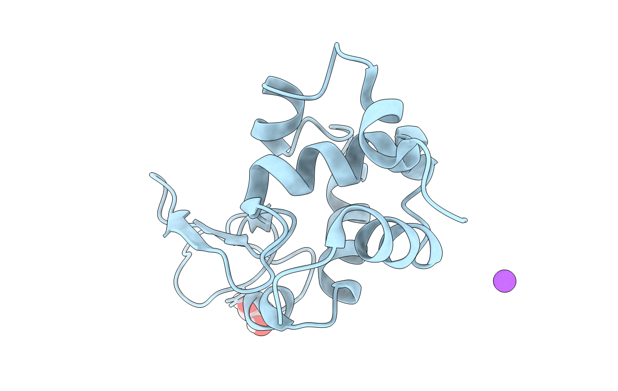 |
Organism: Gallus gallus
Method: X-RAY DIFFRACTION Resolution:3.20 Å Release Date: 2022-07-20 Classification: HYDROLASE Ligands: EDO, NA |
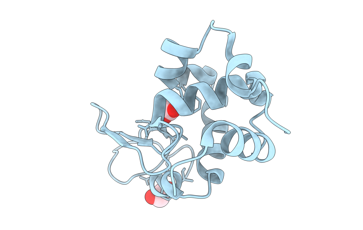 |
Organism: Gallus gallus
Method: X-RAY DIFFRACTION Resolution:2.10 Å Release Date: 2021-10-13 Classification: HYDROLASE Ligands: ACT, EDO, CL |
 |
Organism: Gallus gallus
Method: X-RAY DIFFRACTION Resolution:2.10 Å Release Date: 2021-10-13 Classification: HYDROLASE Ligands: CL, EDO, ACT |
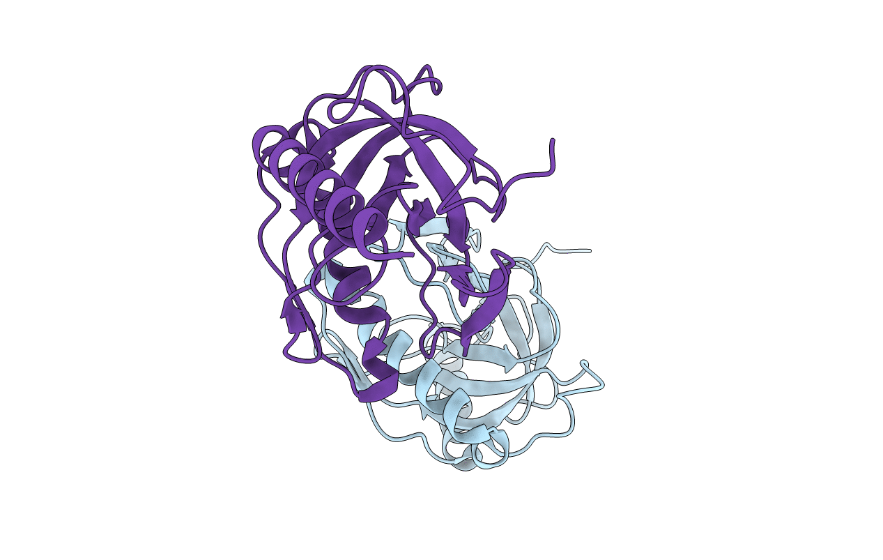 |
Crystal Structure Of Inorganic Pyrophosphatase From Medicago Truncatula (R3 Crystal Form)
Organism: Medicago truncatula
Method: X-RAY DIFFRACTION Resolution:1.84 Å Release Date: 2019-08-14 Classification: HYDROLASE |
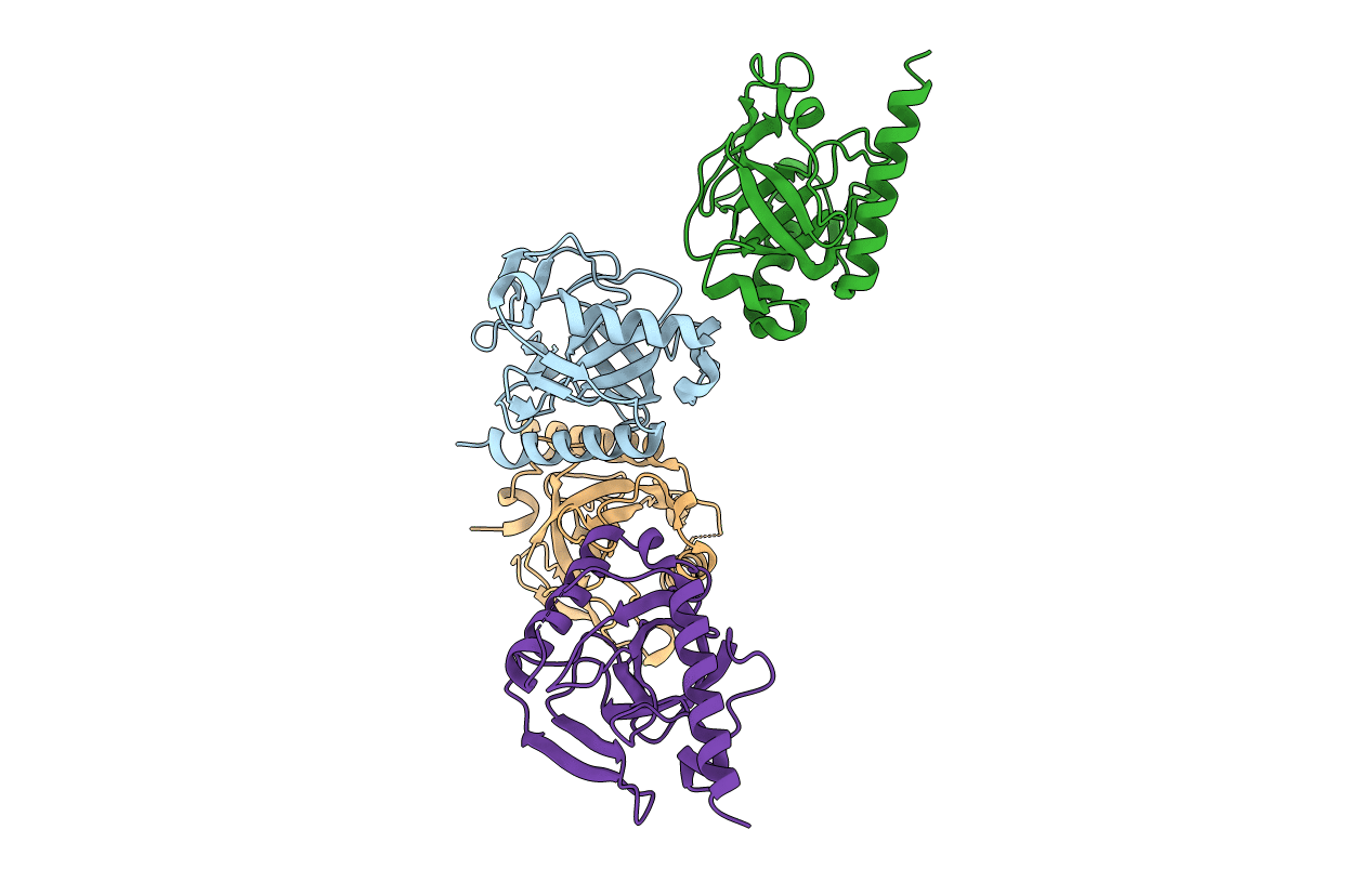 |
Crystal Structure Of Inorganic Pyrophosphatase From Medicago Truncatula (I23 Crystal Form)
Organism: Medicago truncatula
Method: X-RAY DIFFRACTION Resolution:2.89 Å Release Date: 2019-08-14 Classification: HYDROLASE |
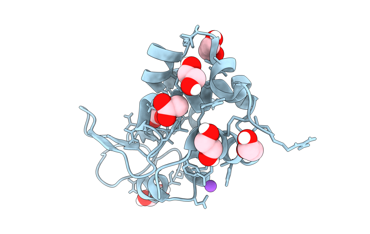 |
Organism: Gallus gallus
Method: X-RAY DIFFRACTION Resolution:1.76 Å Release Date: 2018-10-10 Classification: HYDROLASE Ligands: CL, NA, ACT, EDO |
 |
Organism: Klebsiella pneumoniae
Method: X-RAY DIFFRACTION Resolution:1.69 Å Release Date: 2018-10-10 Classification: ANTIBIOTIC Ligands: NXL |
 |
Crystal Structure Of Yellow Lupin Llpr-10.2B Protein In Complex With Melatonin
Organism: Lupinus luteus
Method: X-RAY DIFFRACTION Resolution:1.51 Å Release Date: 2018-04-18 Classification: PLANT PROTEIN Ligands: UNL, ML1, NA |
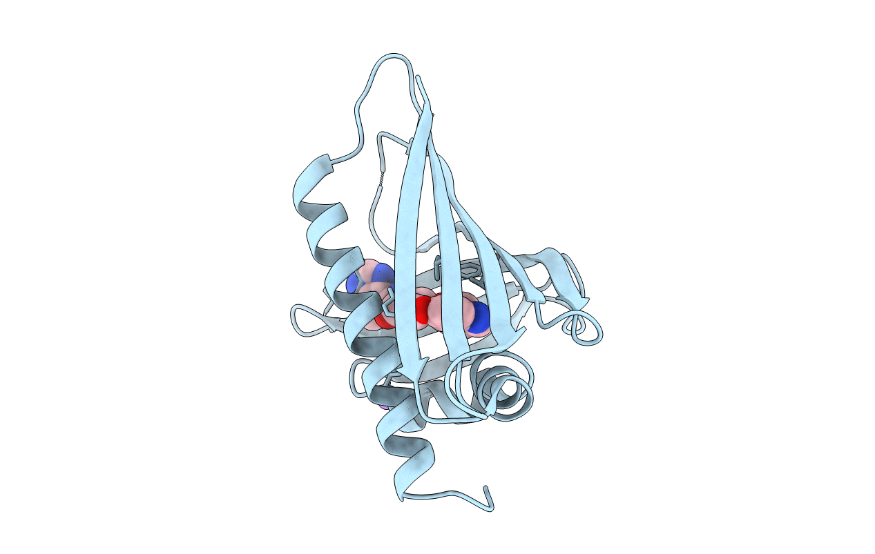 |
Crystal Structure Of Yellow Lupin Llpr-10.2B Protein In Complex With Melatonin And Trans-Zeatin.
Organism: Lupinus luteus
Method: X-RAY DIFFRACTION Resolution:1.57 Å Release Date: 2018-04-18 Classification: PLANT PROTEIN Ligands: UNL, NA, ML1, ZEA |
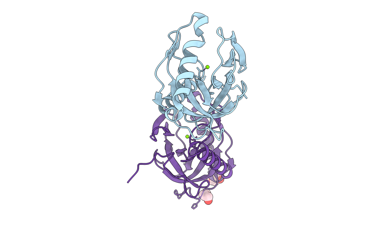 |
Crystal Structure Of Inorganic Pyrophosphatase Ppa1 From Arabidopsis Thaliana
Organism: Arabidopsis thaliana
Method: X-RAY DIFFRACTION Resolution:1.83 Å Release Date: 2017-09-13 Classification: HYDROLASE Ligands: MG, PEG |

