
Search Count: 23
 |
Organism: Micromonospora
Method: X-RAY DIFFRACTION Resolution:2.10 Å Release Date: 2024-08-07 Classification: BIOSYNTHETIC PROTEIN Ligands: GOL |
 |
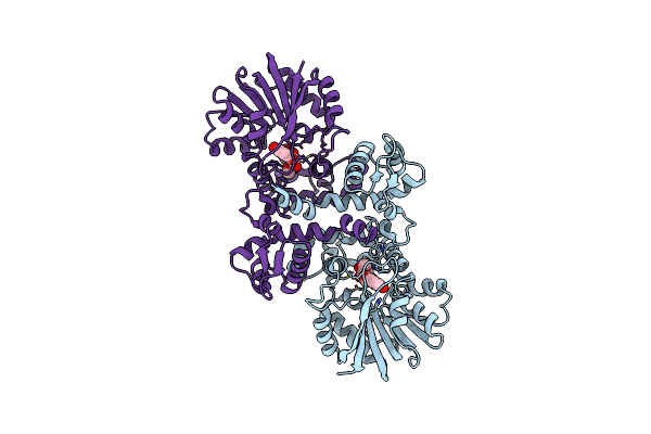
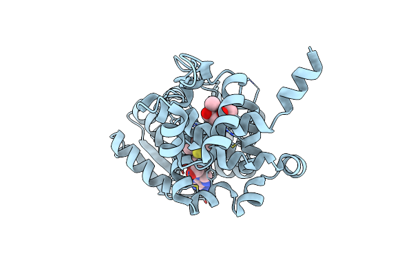
X-Ray Crystallographic Structure Of Snoal2 In Complex With The Polyketide Substrate
Organism: Streptomyces nogalater
Method: X-RAY DIFFRACTION Resolution:1.80 Å Release Date: 2024-08-07 Classification: BIOSYNTHETIC PROTEIN Ligands: XN0 |
 |
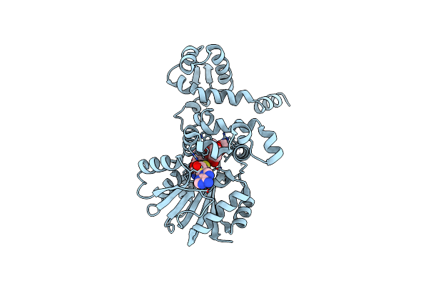
X-Ray Crystallographic Structure Of Snoal2 In Complex With The Polyketide Reaction Product
Organism: Streptomyces nogalater
Method: X-RAY DIFFRACTION Resolution:2.00 Å Release Date: 2024-08-07 Classification: BIOSYNTHETIC PROTEIN Ligands: XN8 |
 |
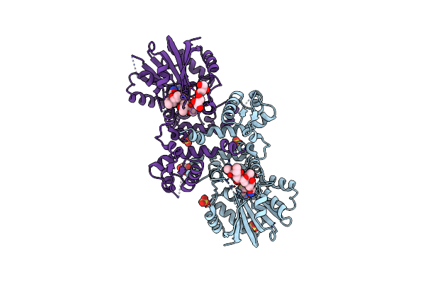
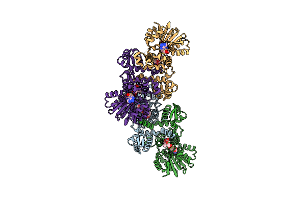
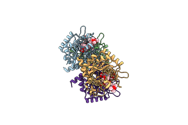
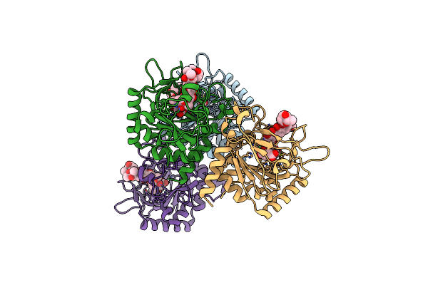
X-Ray Crystallographic Structure Of Swaq2 In Complex With Nadp+ And Doxorubicin
Organism: Streptomyces swartbergensis
Method: X-RAY DIFFRACTION Resolution:2.40 Å Release Date: 2024-08-07 Classification: BIOSYNTHETIC PROTEIN Ligands: DM2, NAP |
 |
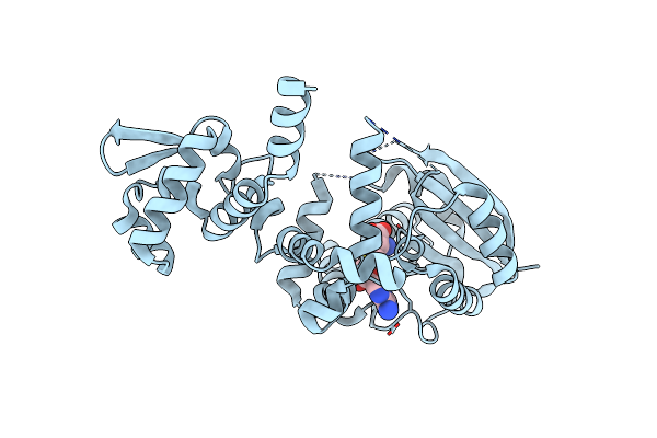
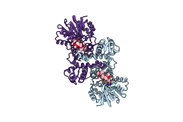
Chimeric Carminomycin-4-O-Methyltransferase (Dnrk) With A Region From 10-Decarboxylase Tamk
Organism: Streptomyces peucetius, Streptomyces tsukubensis (strain dsm 42081 / nbrc 108919 / nrrl 18488 / 9993)
Method: X-RAY DIFFRACTION Resolution:1.53 Å Release Date: 2022-09-07 Classification: BIOSYNTHETIC PROTEIN Ligands: SO4, GOL, SAM, 3VL |
 |
Chimeric Carminomycin-4-O-Methyltransferase (Dnrk) With Regions From 10-Hydroxylase Rdmb And 10-Decarboxylase Tamk
Organism: Streptomyces peucetius, Streptomyces tsukubensis (strain dsm 42081 / nbrc 108919 / nrrl 18488 / 9993), Streptomyces purpurascens
Method: X-RAY DIFFRACTION Resolution:2.32 Å Release Date: 2022-09-07 Classification: BIOSYNTHETIC PROTEIN Ligands: VAK |
 |
Chimeric Carminomycin-4-O-Methyltransferase (Dnrk) With Regions From 10-Hydroxylase Rdmb And 10-Decarboxylase Tamk
Organism: Streptomyces peucetius, Streptomyces tsukubensis (strain dsm 42081 / nbrc 108919 / nrrl 18488 / 9993), Streptomyces purpurascens
Method: X-RAY DIFFRACTION Release Date: 2022-09-07 Classification: BIOSYNTHETIC PROTEIN Ligands: SAH, VAK |
 |
Organism: Streptomyces tsukubensis
Method: X-RAY DIFFRACTION Release Date: 2022-08-24 Classification: BIOSYNTHETIC PROTEIN Ligands: SAH |
 |
Chimeric Carminomycin-4-O-Methyltransferase (Dnrk) With Regions From 10-Hydroxylase Rdmb And 10-Decarboxylase Tamk
Organism: Streptomyces peucetius, Streptomyces tsukubensis (strain dsm 42081 / nbrc 108919 / nrrl 18488 / 9993), Streptomyces purpurascens
Method: X-RAY DIFFRACTION Release Date: 2022-08-24 Classification: BIOSYNTHETIC PROTEIN Ligands: SAH, VAK |
 |
Chimeric Carminomycin-4-O-Methyltransferase (Dnrk) With Regions From 10-Decarboxylate Tamk And 10-Hydroxylase Rdmb, Together With A Single Point Mutation F297G
Organism: Streptomyces peucetius, Streptomyces tsukubensis (strain dsm 42081 / nbrc 108919 / nrrl 18488 / 9993), Streptomyces purpurascens
Method: X-RAY DIFFRACTION Release Date: 2022-08-24 Classification: BIOSYNTHETIC PROTEIN Ligands: SAH, ZBX |
 |
Chimeric Carminomycin-4-O-Methyltransferase (Dnrk) With A Region From 10-Hydroxylase Calmb
Organism: Streptomyces peucetius, Streptomyces sp. zea17i
Method: X-RAY DIFFRACTION Release Date: 2022-07-13 Classification: BIOSYNTHETIC PROTEIN Ligands: 3VL, SAH |
 |
Organism: Streptomyces peucetius, Streptomyces sp. zea17i, Streptomyces purpurascens, Streptomyces tsukubensis (strain dsm 42081 / nbrc 108919 / nrrl 18488 / 9993)
Method: X-RAY DIFFRACTION Resolution:2.39 Å Release Date: 2022-07-13 Classification: BIOSYNTHETIC PROTEIN Ligands: SAH, 3VL |
 |
Organism: Catenulispora acidiphila (strain dsm 44928 / nrrl b-24433 / nbrc 102108 / jcm 14897)
Method: X-RAY DIFFRACTION Resolution:2.50 Å Release Date: 2020-06-17 Classification: UNKNOWN FUNCTION Ligands: GOL, PGE, PG4 |
 |
Organism: Streptomyces sp. cm020
Method: X-RAY DIFFRACTION Resolution:1.80 Å Release Date: 2020-06-17 Classification: ANTIBIOTIC Ligands: GOL |
 |
2.35 Angstrom Structure Of Caci_6494 From Catenulispora Acidiphila In Complex With S-Dnpa
Organism: Catenulispora acidiphila (strain dsm 44928 / nrrl b-24433 / nbrc 102108 / jcm 14897)
Method: X-RAY DIFFRACTION Resolution:2.35 Å Release Date: 2020-06-17 Classification: UNKNOWN FUNCTION Ligands: SDN |
 |
Organism: Streptomyces coelicolor (strain atcc baa-471 / a3(2) / m145)
Method: X-RAY DIFFRACTION Resolution:2.11 Å Release Date: 2020-06-03 Classification: OXIDOREDUCTASE |
 |
Crystal Structure Of The Non-Heme Alpha Ketoglutarate Dependent Epimerase Snon From Nogalamycin Biosynthesis
Organism: Streptomyces nogalater
Method: X-RAY DIFFRACTION Resolution:2.13 Å Release Date: 2016-05-11 Classification: METAL BINDING PROTEIN Ligands: AKG, FE, ACT |
 |
Crystal Structure Of Non-Heme Alpha Ketoglutarate Dependent Carbocyclase Snok From Nogalamycin Biosynthesis
Organism: Streptomyces nogalater
Method: X-RAY DIFFRACTION Resolution:2.24 Å Release Date: 2016-05-11 Classification: LYASE Ligands: FE, AKG, MG |
 |
Crystal Structure Of The Epimerase Snon In Complex With Fe3+, Alpha Ketoglutarate And Nogalamycin Ro
Organism: Streptomyces nogalater
Method: X-RAY DIFFRACTION Resolution:2.20 Å Release Date: 2016-05-11 Classification: ISOMERASE Ligands: FE, AKG, 5R6 |
 |
Crystal Structure Of The Epimerase Snon In Complex With Ni2+, Succinate And Nogalamycin Ro
Organism: Streptomyces nogalater
Method: X-RAY DIFFRACTION Resolution:2.85 Å Release Date: 2016-05-11 Classification: ISOMERASE Ligands: NI, SIN, 5R6 |