
Search Count: 18
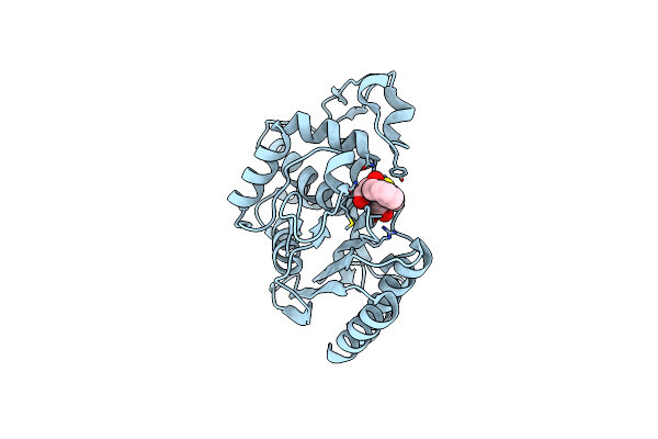 |
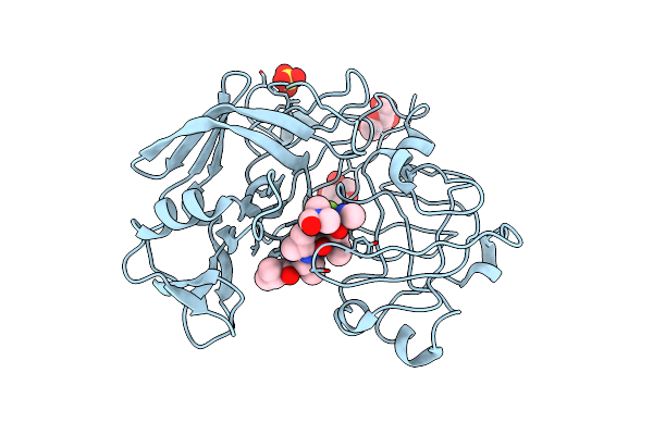
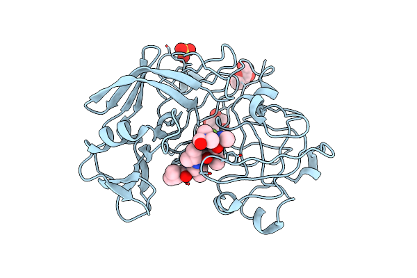
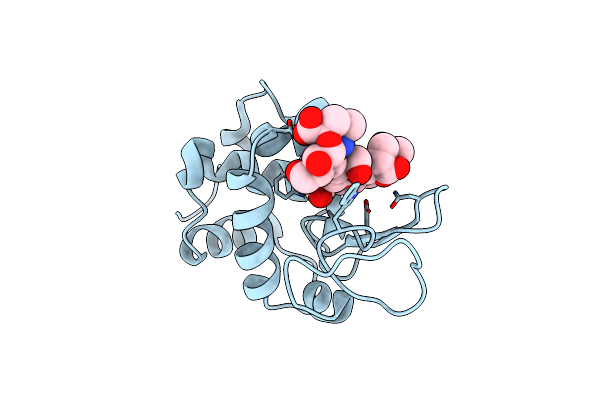
Molecular Structure Of The Acyl-Enzyme Intermediate In Tem-1 Beta-Lactamase
Organism: Escherichia coli
Method: X-RAY DIFFRACTION Resolution:1.70 Å Release Date: 2000-11-01 Classification: HYDROLASE Ligands: PNM |
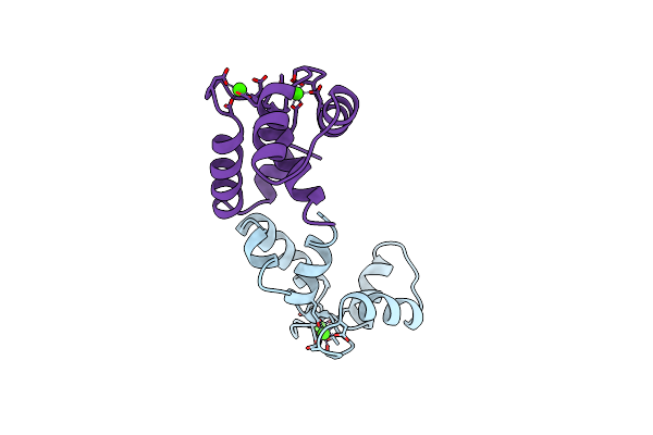 |
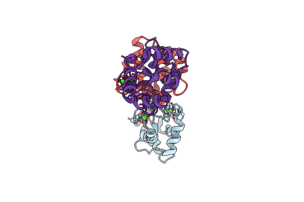
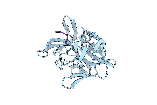
X-Ray Crystallographic Study Of Calcium-Saturated N-Terminal Domain Of Troponin C
Organism: Gallus gallus
Method: X-RAY DIFFRACTION Resolution:1.75 Å Release Date: 1997-12-24 Classification: MUSCLE CONTRACTION Ligands: CA |
 |
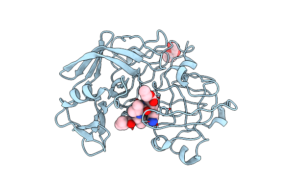
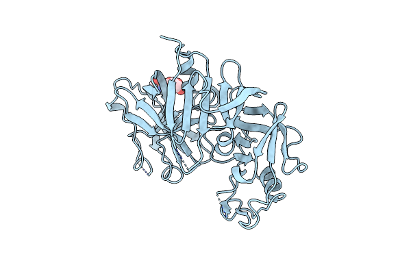
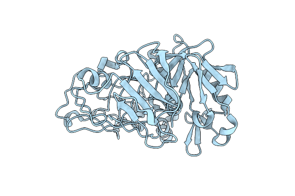
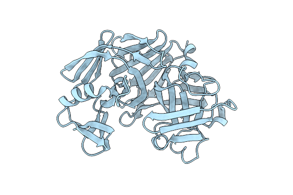
Crystal And Molecular Structures Of Human Progastricsin At 1.62 Angstroms Resolution
Organism: Homo sapiens
Method: X-RAY DIFFRACTION Resolution:1.62 Å Release Date: 1995-01-26 Classification: ASPARTYL PROTEASE |
 |
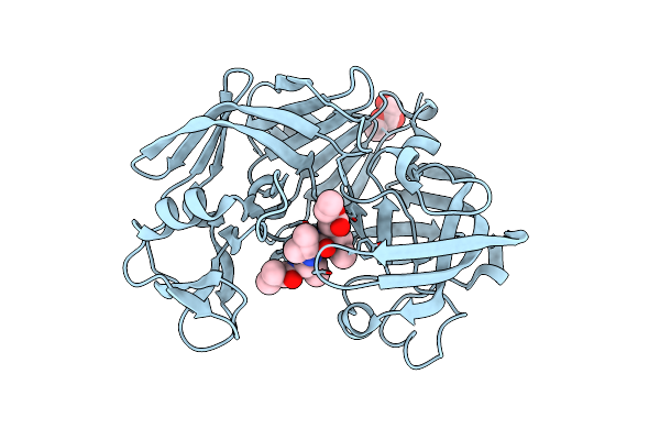
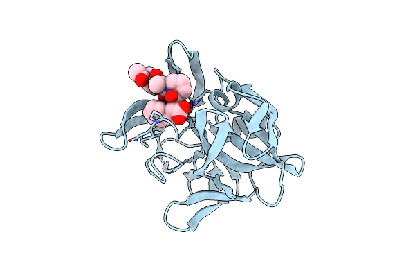
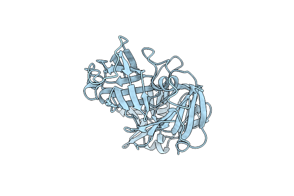
Crystallographic Analysis Of A Pepstatin Analogue Binding To The Aspartyl Proteinase Penicillopepsin At 1.8 Angstroms Resolution
Organism: Penicillium janthinellum
Method: X-RAY DIFFRACTION Resolution:1.80 Å Release Date: 1994-01-31 Classification: HYDROLASE/HYDROLASE INHIBITOR Ligands: MAN |
 |
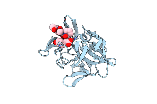
Crystallographic Analysis Of A Pepstatin Analogue Binding To The Aspartyl Proteinase Penicillopepsin At 1.8 Angstroms Resolution
Organism: Penicillium janthinellum
Method: X-RAY DIFFRACTION Resolution:1.80 Å Release Date: 1994-01-31 Classification: HYDROLASE/HYDROLASE INHIBITOR Ligands: MAN |
 |
Crystallographic Analysis Of Transition State Mimics Bound To Penicillopepsin: Difluorostatine-And Difluorostatone-Containing Peptides
Organism: Penicillium janthinellum
Method: X-RAY DIFFRACTION Resolution:1.80 Å Release Date: 1994-01-31 Classification: HYDROLASE/HYDROLASE INHIBITOR Ligands: MAN, XYS, SO4, DMF |
 |
Crystallographic Analysis Of Transition State Mimics Bound To Penicillopepsin: Difluorostatine-And Difluorostatone-Containing Peptides
Organism: Penicillium janthinellum
Method: X-RAY DIFFRACTION Resolution:1.80 Å Release Date: 1994-01-31 Classification: HYDROLASE/HYDROLASE INHIBITOR Ligands: MAN, HSY, SO4, DMF |
 |
Refined X-Ray Structure Of Rat Parvalbumin, A Mammalian Alpha-Lineage Parvalbumin, At 2.0 A Resolution
Organism: Rattus rattus
Method: X-RAY DIFFRACTION Resolution:2.00 Å Release Date: 1994-01-31 Classification: CALCIUM-BINDING PROTEIN Ligands: CA |
 |
Structure Of Recombinant Human Renin, A Target For Cardiovascular-Active Drugs, At 2.5 Angstroms Resolution
Organism: Homo sapiens
Method: X-RAY DIFFRACTION Resolution:2.50 Å Release Date: 1994-01-31 Classification: HYDROLASE(ACID PROTEINASE) Ligands: NAG |
 |
Organism: Sus scrofa
Method: X-RAY DIFFRACTION Resolution:1.80 Å Release Date: 1992-10-15 Classification: HYDROLASE(ACID PROTEINASE ZYMOGEN) |
 |
Structures Of Product And Inhibitor Complexes Of Streptomyces Griseus Protease A At 1.8 Angstroms Resolution. A Model For Serine Protease Catalysis
Organism: Streptomyces griseus
Method: X-RAY DIFFRACTION Resolution:1.80 Å Release Date: 1991-10-15 Classification: HYDROLASE/HYDROLASE INHIBITOR |
 |
Structures Of Product And Inhibitor Complexes Of Streptomyces Griseus Protease A At 1.8 Angstroms Resolution. A Model For Serine Protease Catalysis
Organism: Streptomyces griseus
Method: X-RAY DIFFRACTION Resolution:1.80 Å Release Date: 1991-10-15 Classification: HYDROLASE/HYDROLASE INHIBITOR |
 |
Structures Of Product And Inhibitor Complexes Of Streptomyces Griseus Protease A At 1.8 Angstroms Resolution. A Model For Serine Protease Catalysis
Organism: Streptomyces griseus
Method: X-RAY DIFFRACTION Resolution:1.80 Å Release Date: 1991-10-15 Classification: HYDROLASE/HYDROLASE INHIBITOR |
 |
Organism: Penicillium janthinellum
Method: X-RAY DIFFRACTION Resolution:1.80 Å Release Date: 1991-01-15 Classification: HYDROLASE (ACID PROTEINASE) |
 |
The Molecular And Crystal Structures Of Monoclinic Porcine Pepsin Refined At 1.8 Angstroms Resolution
Organism: Sus scrofa
Method: X-RAY DIFFRACTION Resolution:1.80 Å Release Date: 1990-04-15 Classification: HYDROLASE (ACID PROTEINASE) |
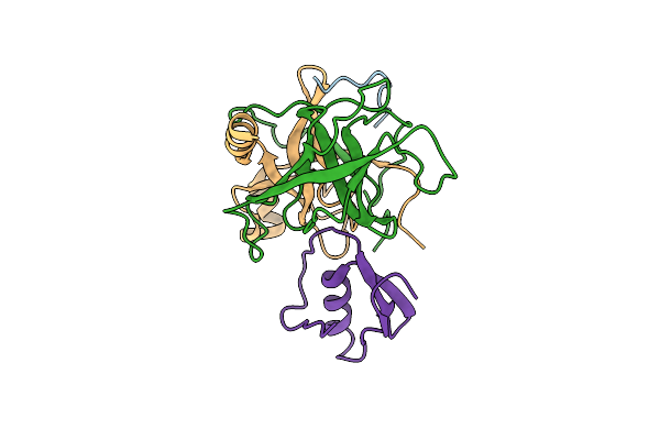 |
Crystal And Molecular Structures Of The Complex Of Alpha-*Chymotrypsin With Its Inhibitor Turkey Ovomucoid Third Domain At 1.8 Angstroms Resolution
Organism: Bos taurus, Meleagris gallopavo
Method: X-RAY DIFFRACTION Resolution:1.80 Å Release Date: 1988-07-16 Classification: HYDROLASE/HYDROLASE INHIBITOR |
 |
Structure Of The Complex Of Streptomyces Griseus Protease B And The Third Domain Of The Turkey Ovomucoid Inhibitor At 1.8 Angstroms Resolution
Organism: Streptomyces griseus
Method: X-RAY DIFFRACTION Resolution:1.80 Å Release Date: 1983-07-12 Classification: COMPLEX(SERINE PROTEINASE-INHIBITOR) |
 |
X-Ray Crystallography Of The Binding Of The Bacterial Cell Wall Trisaccharide Nam-Nag-Nam To Lysozyme
Organism: Gallus gallus
Method: X-RAY DIFFRACTION Resolution:2.50 Å Release Date: 1980-02-27 Classification: HYDROLASE (O-GLYCOSYL) |