
Search Count: 16
 |
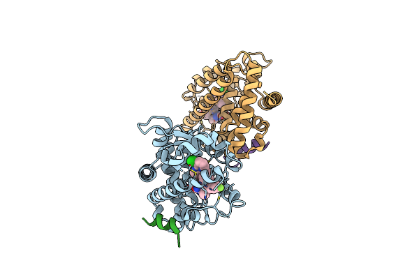
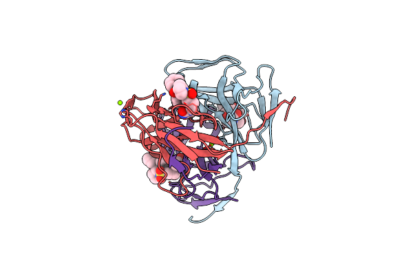
Crystal Structure Of Rorgt With Compound (4R)-6-[(2,5-Dichloro-3-{[(2R,4R)-1-(Cyclopentanecarbonyl)-2-Methylpiperidin-4-Yl]Oxy}Phenyl)Amino]-6-Oxo-4-Phenylhexanoic Acid
Organism: Homo sapiens
Method: X-RAY DIFFRACTION Resolution:1.90 Å Release Date: 2021-02-03 Classification: NUCLEAR PROTEIN Ligands: VK4, GOL, SO4 |
 |
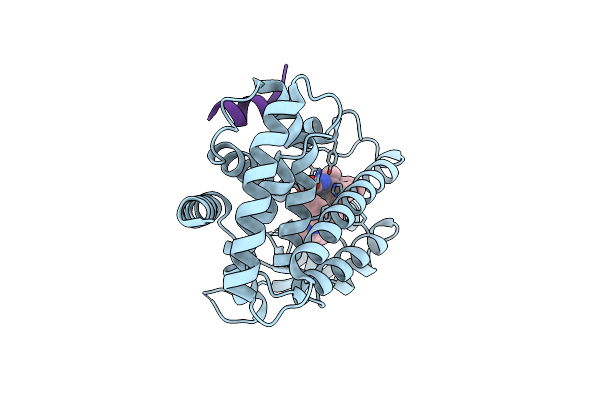
Crystal Structure Of Rorgt With 3-Cyano-N-(3-{[(3S)-4-(Cyclopentanecarbonyl)-3-Methylpiperazin-1-Yl]Methyl}-5-Fluoro-2-Methylphenyl)Benzamide (Compound 1)
Organism: Homo sapiens
Method: X-RAY DIFFRACTION Resolution:2.40 Å Release Date: 2020-03-04 Classification: PROTEIN BINDING Ligands: LKY, EDO |
 |
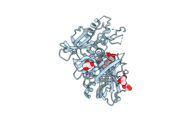
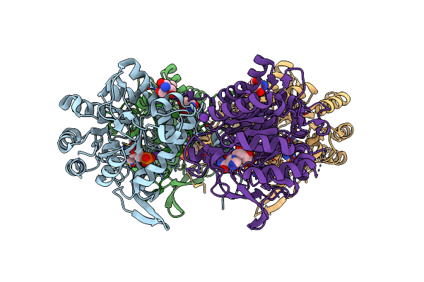
Crystal Structure Of Ppargamma Ligand Binding Domain In Complex With Dibenzooxepine Derivative Compound-17
Organism: Homo sapiens
Method: X-RAY DIFFRACTION Resolution:1.84 Å Release Date: 2019-10-30 Classification: NUCLEAR PROTEIN Ligands: CTU |
 |
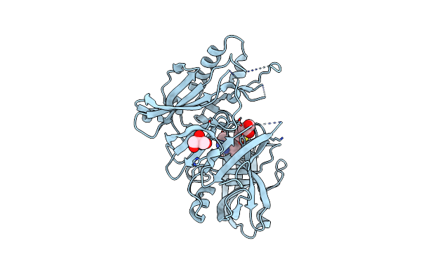
Crystal Structure Of Ppargamma Ligand Binding Domain In Complex With Dibenzooxepine Derivative Compound-9
Organism: Homo sapiens
Method: X-RAY DIFFRACTION Resolution:2.20 Å Release Date: 2018-11-14 Classification: NUCLEAR PROTEIN Ligands: KK4 |
 |
Organism: Homo sapiens
Method: X-RAY DIFFRACTION Release Date: 2017-09-13 Classification: TRANSFERASE/TRANSFERASE INHIBITOR Ligands: GF5, MES |
 |
Crystal Structure Of Beta Secetase In Complex With 2-Amino-3,6-Dimethyl-6-(2-Phenylethyl)-3,4,5,6-Tetrahydropyrimidin-4-One
Organism: Homo sapiens
Method: X-RAY DIFFRACTION Resolution:2.25 Å Release Date: 2013-10-02 Classification: HYDROLASE Ligands: GOL, IOD, 0B3 |
 |
Crystal Structure Of Beta Secetase In Complex With (6S)-2-Amino-3,6-Dimethyl-6-[(1R,2R)-2-Phenylcyclopropyl]-3,4,5,6-Tetrahydropyrimidin-4-One
Organism: Homo sapiens
Method: X-RAY DIFFRACTION Resolution:2.50 Å Release Date: 2013-10-02 Classification: HYDROLASE Ligands: IOD, GOL, DMS, 0B4 |
 |
Organism: Saccharomyces cerevisiae
Method: X-RAY DIFFRACTION Resolution:2.80 Å Release Date: 2013-04-17 Classification: HYDROLASE/HYDROLASE INHIBITOR Ligands: 1KR |
 |
Crystal Structure Of Beta Secetase In Complex With 2-Amino-3-Methyl-6-((1S, 2R)-2-Phenylcyclopropyl)Pyrimidin-4(3H)-One
Organism: Homo sapiens
Method: X-RAY DIFFRACTION Resolution:2.05 Å Release Date: 2012-10-24 Classification: HYDROLASE/HYDROLASE INHIBITOR Ligands: IOD, GOL, B00 |
 |
Crystal Structure Of Beta Secetase In Complex With 2-Amino-6-((1S,2R)-2-(3'-Methoxybiphenyl-3-Yl)Cyclopropyl)-3-Methylpyrimidin-4(3H)-One
Organism: Homo sapiens
Method: X-RAY DIFFRACTION Resolution:2.10 Å Release Date: 2012-10-24 Classification: HYDROLASE/HYDROLASE INHIBITOR Ligands: IOD, GOL, 0B1 |
 |
Crystal Structure Of Beta Secetase In Complex With 2-Amino-3-Methyl-6-((1S,2R)-2-(3'-Methylbiphenyl-4-Yl)Cyclopropyl)Pyrimidin-4(3H)-One
Organism: Homo sapiens
Method: X-RAY DIFFRACTION Resolution:2.50 Å Release Date: 2012-10-24 Classification: HYDROLASE/HYDROLASE INHIBITOR Ligands: GOL, B02 |
 |
Cd38 Structure-Based Inhibitor Design Using The N1-Cyclic Inosine 5'-Diphosphate Ribose Template
Organism: Homo sapiens
Method: X-RAY DIFFRACTION Resolution:1.88 Å Release Date: 2012-10-10 Classification: HYDROLASE/HYDROLASE INHIBITOR Ligands: C8R |
 |
Cd38 Structure-Based Inhibitor Design Using The N1-Cyclic Inosine 5'-Diphosphate Ribose Template
Organism: Homo sapiens
Method: X-RAY DIFFRACTION Resolution:2.12 Å Release Date: 2012-10-10 Classification: HYDROLASE/HYDROLASE INHIBITOR Ligands: CVR |
 |
Organism: Homo sapiens
Method: X-RAY DIFFRACTION Resolution:1.80 Å Release Date: 2010-12-15 Classification: Hydrolase/Hydrolase inhibitor Ligands: MSJ, MG |
 |
Organism: Homo sapiens
Method: X-RAY DIFFRACTION Resolution:1.70 Å Release Date: 2010-12-08 Classification: Hydrolase/Hydrolase inhibitor Ligands: MKH, MG |
 |
The Crystal Structure Of The Complex Between Imp Dehydrogenase Catalytic Domain And A Transition State Analogue Mzp
Organism: Tritrichomonas foetus
Method: X-RAY DIFFRACTION Resolution:2.00 Å Release Date: 2003-07-22 Classification: OXIDOREDUCTASE Ligands: K, MZP, TRS |