
Search Count: 6
 |
Organism: Homo sapiens
Method: X-RAY DIFFRACTION Resolution:2.75 Å Release Date: 2019-09-11 Classification: REPLICATION Ligands: ZN, MN, ATP |
 |
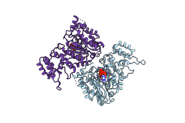
Crystal Structure Of The Pri1 Subunit Of Human Primase Bound To Vidarabine Triphosphate
Organism: Homo sapiens
Method: X-RAY DIFFRACTION Resolution:2.35 Å Release Date: 2019-09-11 Classification: REPLICATION Ligands: ZN, MN, HEJ, EDO |
 |
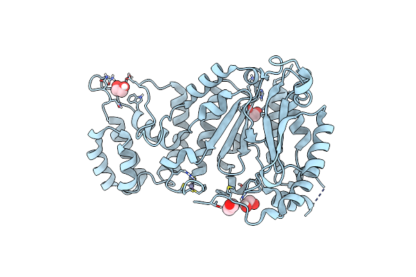
Crystal Structure Of The Pri1 Subunit Of Human Primase Bound To Fludarabine Triphosphate
Organism: Homo sapiens
Method: X-RAY DIFFRACTION Resolution:2.20 Å Release Date: 2019-09-11 Classification: REPLICATION Ligands: ZN, MN, HFD, EDO |
 |
Organism: Homo sapiens
Method: X-RAY DIFFRACTION Resolution:1.95 Å Release Date: 2019-09-11 Classification: REPLICATION Ligands: ZN, MN, DTP, EDO |
 |
Organism: Homo sapiens
Method: X-RAY DIFFRACTION Resolution:1.85 Å Release Date: 2019-09-11 Classification: REPLICATION Ligands: ZN, MN, JSQ, EDO |
 |
Organism: Homo sapiens
Method: X-RAY DIFFRACTION Resolution:1.50 Å Release Date: 2019-09-11 Classification: REPLICATION Ligands: EDO, ZN |