
Search Count: 29
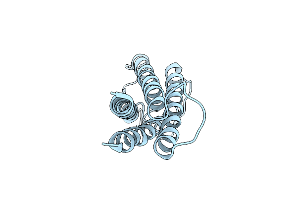 |
Organism: Homo sapiens
Method: ELECTRON CRYSTALLOGRAPHY Resolution:3.50 Å Release Date: 2025-05-21 Classification: CELL ADHESION |
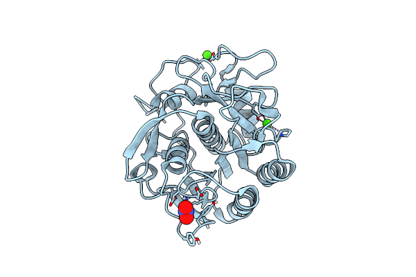 |
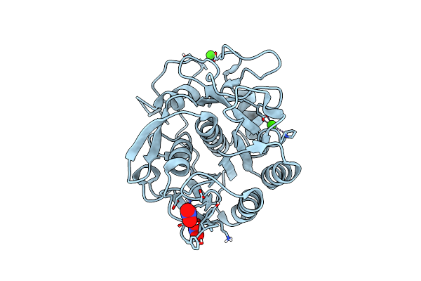
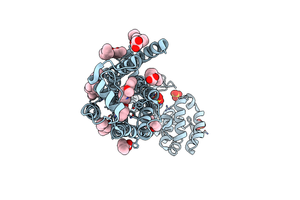
Microed Structure Of Proteinase K From Lamellae Milled From Multiple Plasma Sources
Organism: Parengyodontium album
Method: ELECTRON CRYSTALLOGRAPHY Resolution:1.39 Å Release Date: 2023-05-24 Classification: HYDROLASE Ligands: NO3, CA |
 |
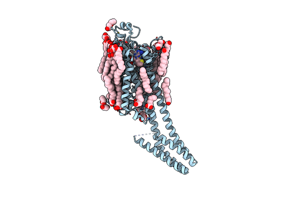
Organism: Homo sapiens
Method: ELECTRON CRYSTALLOGRAPHY Resolution:2.00 Å Release Date: 2023-03-08 Classification: MEMBRANE PROTEIN Ligands: ZMA, CLR, OLA, OLC, OLB, NA |
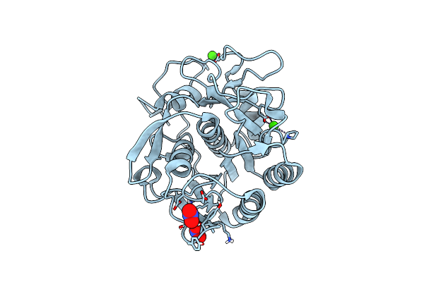 |
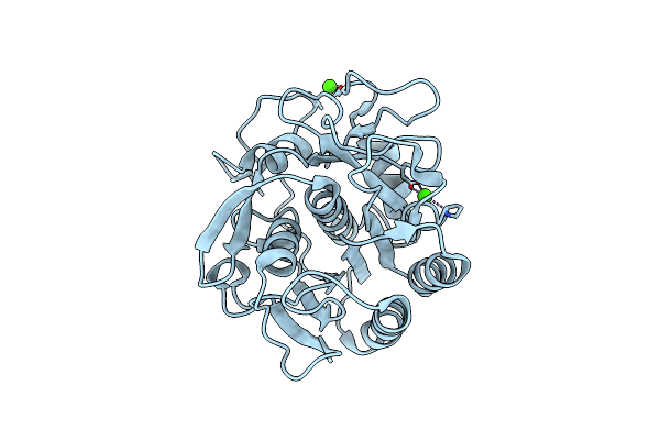
Organism: Parengyodontium album
Method: ELECTRON CRYSTALLOGRAPHY Resolution:1.45 Å Release Date: 2023-03-08 Classification: HYDROLASE Ligands: NO3, CA |
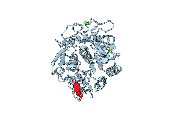 |
Organism: Parengyodontium album
Method: ELECTRON CRYSTALLOGRAPHY Resolution:1.40 Å Release Date: 2023-03-08 Classification: HYDROLASE Ligands: NO3, CA |
 |
Organism: Parengyodontium album
Method: ELECTRON CRYSTALLOGRAPHY Resolution:1.50 Å Release Date: 2023-03-08 Classification: HYDROLASE Ligands: NO3, CA |
 |
Organism: Parengyodontium album
Method: ELECTRON CRYSTALLOGRAPHY Resolution:1.80 Å Release Date: 2023-03-08 Classification: HYDROLASE Ligands: CA |
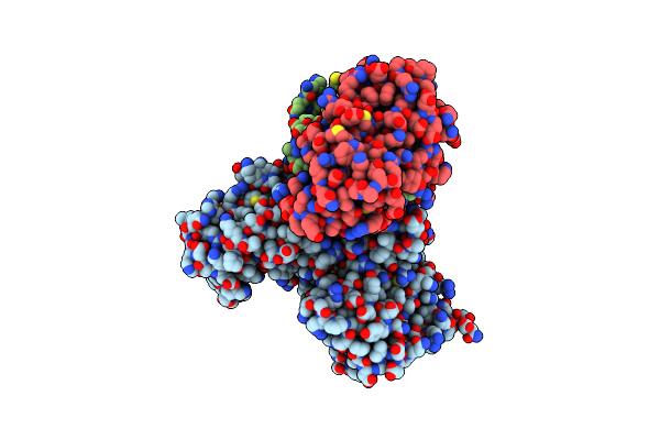 |
Organism: Macaca mulatta, Human immunodeficiency virus 1, Homo sapiens, Escherichia coli
Method: ELECTRON MICROSCOPY Release Date: 2023-01-11 Classification: VIRAL PROTEIN/RNA Ligands: ZN |
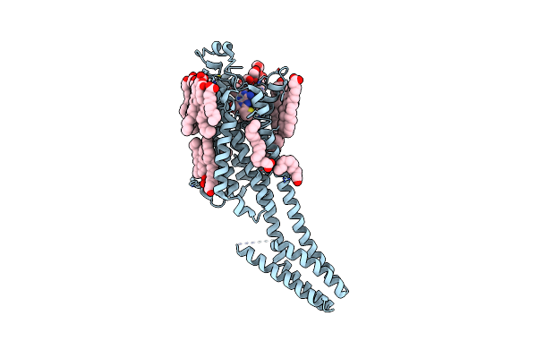 |
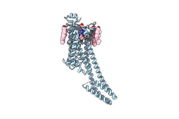
Crystal Structure Of A2Aar-Star2-S277-Bril In Complex With A Novel A2A Antagonist, Lj-4517
Organism: Homo sapiens, Escherichia coli
Method: X-RAY DIFFRACTION Resolution:2.80 Å Release Date: 2022-08-31 Classification: MEMBRANE PROTEIN Ligands: OLA, OLC, PEG, LJX, CLR |
 |
Crystal Structure Of A2Aar-Star2-Bril In Complex With A Novel A2A Antagonist, Lj-4517
Organism: Homo sapiens, Escherichia coli
Method: X-RAY DIFFRACTION Resolution:2.05 Å Release Date: 2022-08-31 Classification: MEMBRANE PROTEIN Ligands: PEG, LJX, NA, OLC, OLA, CLR, ETF |
 |
Organism: Homo sapiens
Method: X-RAY DIFFRACTION Resolution:2.50 Å Release Date: 2022-08-10 Classification: MEMBRANE PROTEIN Ligands: TKO, CLR, OLC, OLA |
 |
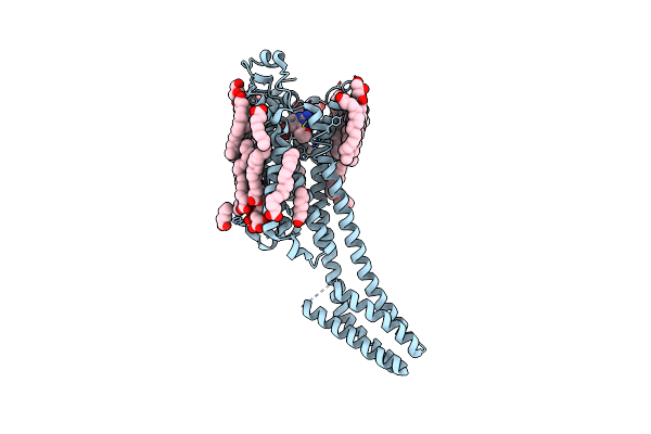
Organism: Homo sapiens, Escherichia coli
Method: ELECTRON CRYSTALLOGRAPHY Resolution:2.79 Å Release Date: 2021-09-08 Classification: MEMBRANE PROTEIN Ligands: ZMA, CLR, NA |
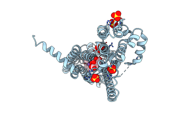 |
Xfel Crystal Structure Of The Prostaglandin D2 Receptor Crth2 In Complex With 15R-Methyl-Pgd2
Organism: Homo sapiens, Enterobacteria phage t4
Method: X-RAY DIFFRACTION Resolution:2.61 Å Release Date: 2021-08-25 Classification: MEMBRANE PROTEIN Ligands: YSS, NA, FLC, SO4 |
 |
Organism: Homo sapiens, Enterobacteria phage t4
Method: X-RAY DIFFRACTION Resolution:2.80 Å Release Date: 2019-11-13 Classification: MEMBRANE PROTEIN Ligands: JTZ, CLR, OLC, OLA, SO4 |
 |
Organism: Homo sapiens, Enterobacteria phage t4
Method: X-RAY DIFFRACTION Resolution:3.40 Å Release Date: 2019-11-13 Classification: MEMBRANE PROTEIN Ligands: CAU, SO4, CLR, OLC, OLA |
 |
Organism: Homo sapiens, Enterobacteria phage t4
Method: X-RAY DIFFRACTION Resolution:3.20 Å Release Date: 2019-11-13 Classification: MEMBRANE PROTEIN Ligands: TIM, SO4, CLR, OLC, OLA |
 |
Organism: Homo sapiens, Enterobacteria phage t4
Method: X-RAY DIFFRACTION Resolution:2.40 Å Release Date: 2019-11-13 Classification: MEMBRANE PROTEIN Ligands: JTZ, CLR, OLB, OLC, SO4, OLA |
 |
Organism: Homo sapiens, Enterobacteria phage t4
Method: X-RAY DIFFRACTION Resolution:2.50 Å Release Date: 2019-11-13 Classification: MEMBRANE PROTEIN Ligands: CVD, SO4, CLR, OLC, OLA |
 |
Organism: Homo sapiens, Enterobacteria phage t4
Method: X-RAY DIFFRACTION Resolution:2.60 Å Release Date: 2019-11-13 Classification: MEMBRANE PROTEIN Ligands: JRZ, SO4, CLR, OLA, OLC |
 |
Organism: Homo sapiens, Enterobacteria phage t4
Method: X-RAY DIFFRACTION Resolution:2.90 Å Release Date: 2019-11-13 Classification: MEMBRANE PROTEIN Ligands: SNP, SO4, CLR, OLA, OLC |