
Search Count: 9
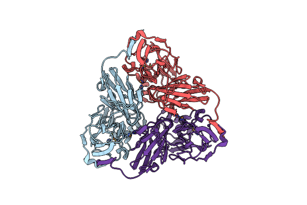 |
Organism: Achromobacter cycloclastes
Method: ELECTRON MICROSCOPY Release Date: 2020-08-12 Classification: OXIDOREDUCTASE Ligands: CU |
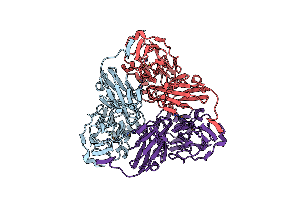 |
Organism: Achromobacter cycloclastes
Method: ELECTRON MICROSCOPY Release Date: 2020-08-12 Classification: OXIDOREDUCTASE Ligands: CU |
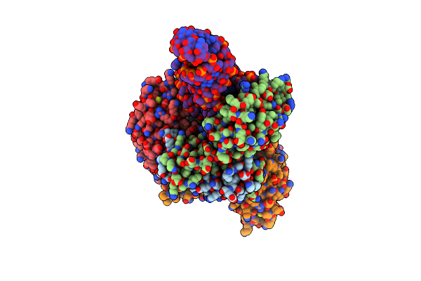 |
Crystal Structure Of Sepcyse-Sepcyss In Complex With Trnacys From Methanocaldococcus Jannaschii
Organism: Methanocaldococcus jannaschii dsm 2661, Methanocaldococcus jannaschii
Method: X-RAY DIFFRACTION Resolution:2.60 Å Release Date: 2017-12-06 Classification: RNA BINDING PROTEIN/RNA |
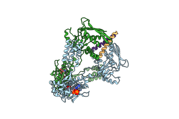 |
Organism: Methanocaldococcus jannaschii dsm 2661, Methanocaldococcus jannaschii
Method: X-RAY DIFFRACTION Resolution:3.10 Å Release Date: 2017-12-06 Classification: RNA BINDING PROTEIN Ligands: ATP, SO4 |
 |
Organism: Pseudomonas aeruginosa pao1
Method: X-RAY DIFFRACTION Resolution:1.65 Å Release Date: 2016-09-28 Classification: HYDROLASE Ligands: MN, LP5, GOL |
 |
Organism: Pseudomonas aeruginosa pao1
Method: X-RAY DIFFRACTION Resolution:1.72 Å Release Date: 2016-09-28 Classification: HYDROLASE Ligands: LP5, GOL |
 |
Organism: Pseudomonas aeruginosa pao1
Method: X-RAY DIFFRACTION Resolution:1.60 Å Release Date: 2016-09-28 Classification: HYDROLASE Ligands: LP5, GOL |
 |
Organism: Pseudomonas aeruginosa pao1
Method: X-RAY DIFFRACTION Resolution:1.96 Å Release Date: 2016-09-28 Classification: HYDROLASE Ligands: MN, GOL |
 |
Organism: Pseudomonas aeruginosa pao1
Method: X-RAY DIFFRACTION Resolution:1.75 Å Release Date: 2016-09-28 Classification: HYDROLASE Ligands: GOL |