
Search Count: 20
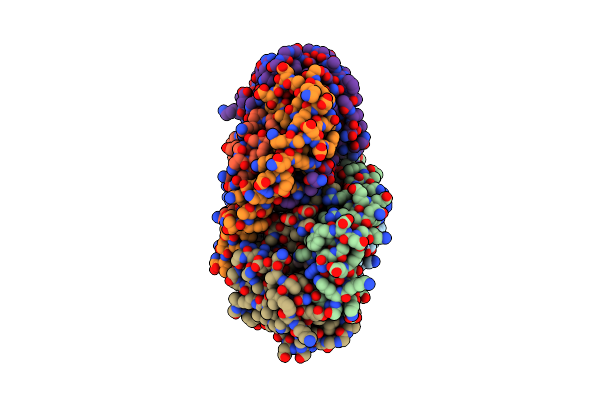 |
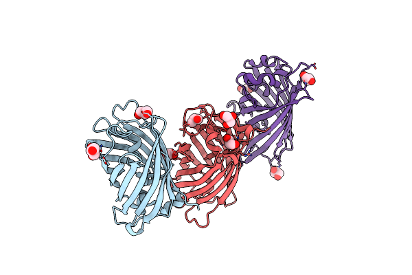
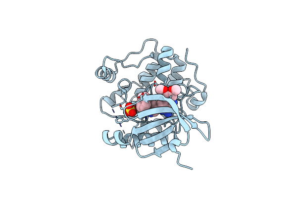
Crystal Structure Of The Soluble Green Pigment Protein From Tettigonia Cantans
Organism: Tettigonia cantans
Method: X-RAY DIFFRACTION Release Date: 2025-04-23 Classification: TRANSPORT PROTEIN Ligands: LUT, AZI, PLC, A1L6M |
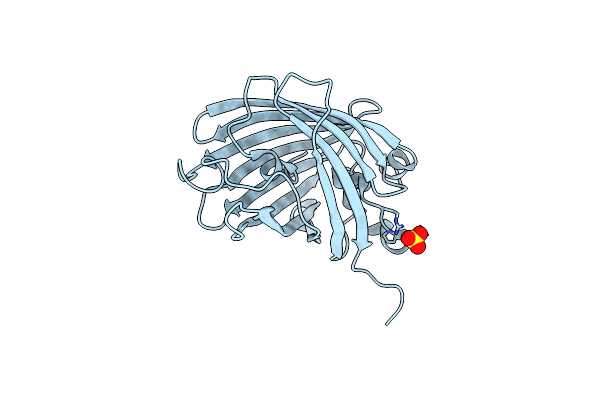 |
Organism: Recombinant vesicular stomatitis indiana virus rvsv-g/gfp
Method: X-RAY DIFFRACTION Resolution:2.20 Å Release Date: 2021-06-16 Classification: FLUORESCENT PROTEIN Ligands: SO4 |
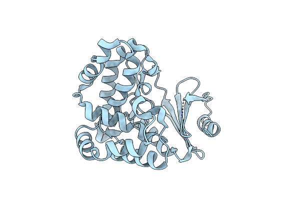 |
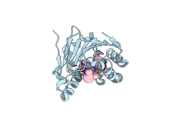
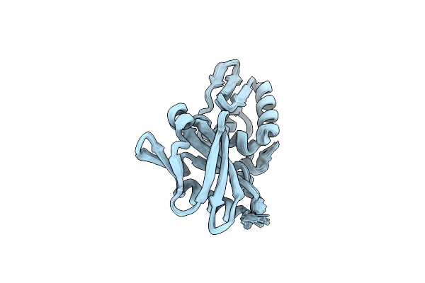
Structure Of Aminoglycoside Phosphotransferase Aph(3'')-Id From Streptomyces Rimosus Atcc10970
Organism: Streptomyces rimosus
Method: X-RAY DIFFRACTION Resolution:1.17 Å Release Date: 2019-03-20 Classification: TRANSFERASE |
 |
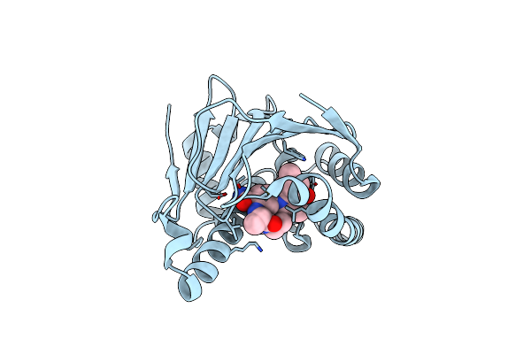
Structure Of Aminoglycoside Phosphotransferase Aph(3'')-Id From Streptomyces Rimosus Atcc10970 In Complex With Adp And Streptomycin
Organism: Streptomyces rimosus subsp. rimosus atcc 10970
Method: X-RAY DIFFRACTION Resolution:1.65 Å Release Date: 2019-03-20 Classification: TRANSFERASE Ligands: GOL, ADP, SRY |
 |
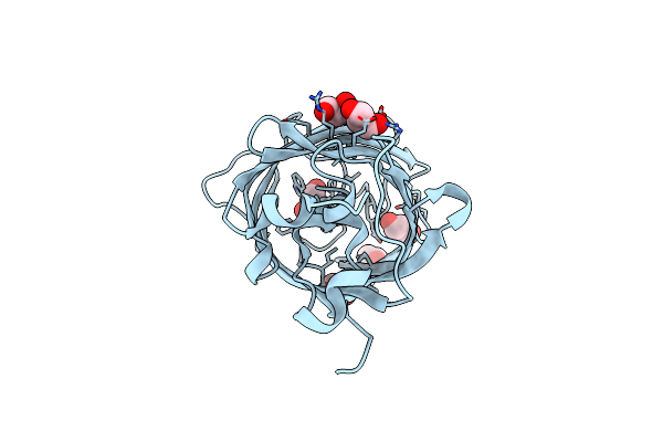
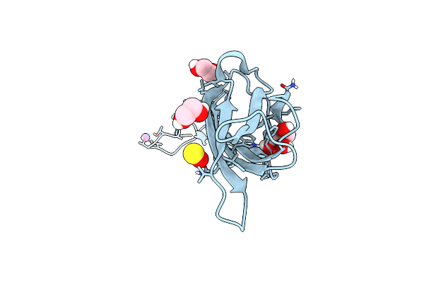
Crystal Structure Of The Green Fluorescent Variant, Nowgfp, Of The Cyan Cerulean At Ph 9.0
Organism: Synthetic
Method: X-RAY DIFFRACTION Resolution:1.35 Å Release Date: 2015-09-02 Classification: FLUORESCENT PROTEIN Ligands: GOL |
 |
Crystal Structure Of The Green Fluorescent Rotein Nowgfp (The Variant Of Cyan Cerulean) At Ph 4.8
Organism: Cfp marker plasmid pwm1009
Method: X-RAY DIFFRACTION Resolution:1.18 Å Release Date: 2015-09-02 Classification: FLUORESCENT PROTEIN Ligands: GOL |
 |
Crystal Structure Of The Photoconverted Green Fluorescent Protein Nowgfp_Conv (The Variant Of Cyan Cerulean) At Ph 7.0
Method: X-RAY DIFFRACTION
Resolution:2.50 Å Release Date: 2015-09-02 Classification: FLUORESCENT PROTEIN Ligands: GOL |
 |
Crystal Structure Of The Tail Fiber Gp53 From Acinetobacter Baumannii Bacteriophage Ap22
Organism: Acinetobacter bacteriophage ap22
Method: X-RAY DIFFRACTION Resolution:1.37 Å Release Date: 2014-10-01 Classification: STRUCTURAL PROTEIN Ligands: GOL, BR, NA, EDO, SO3 |
 |
Organism: Human immunodeficiency virus 1
Method: SOLUTION NMR Release Date: 2011-11-09 Classification: PROTEIN TRANSPORT |
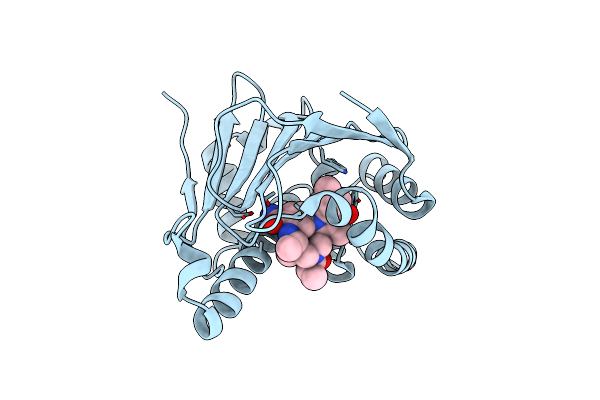 |
Discovery Of A Stable Macrocyclic O-Aminobenzamide Hsp90 Inhibitor Capable Of Significantly Decreasing Tumor Volume In A Mouse Xenograft Model.
Organism: Homo sapiens
Method: X-RAY DIFFRACTION Resolution:1.57 Å Release Date: 2011-07-13 Classification: CHAPERONE/CHAPERONE INHIBITOR Ligands: 06T |
 |
Discovery Of A Macrocyclic O-Aminobenzamide Hsp90 Inhibitor With Heterocyclic Tether That Shows Extended Biomarker Activity And In Vivo Efficacy In A Mouse Xenograft Model.
Organism: Homo sapiens
Method: X-RAY DIFFRACTION Resolution:1.58 Å Release Date: 2011-06-08 Classification: CHAPERONE/CHAPERONE INHIBITOR Ligands: 06J |
 |
Macrocyclic Lactams As Potent Hsp90 Inhibitors With Excellent Tumor Exposure And Extended Biomarker Activity.
Organism: Homo sapiens
Method: X-RAY DIFFRACTION Resolution:1.58 Å Release Date: 2011-05-11 Classification: CHAPERONE/CHAPERONE INHIBITOR Ligands: 06H |
 |
Design And Sar Of Macrocyclic Hsp90 Inhibitors With Increased Metabolic Stability And Potent Cell-Proliferation Activity
Organism: Homo sapiens
Method: X-RAY DIFFRACTION Resolution:1.57 Å Release Date: 2011-04-06 Classification: CHAPERONE/CHAPERONE INHIBITOR Ligands: 05S, DMS |
 |
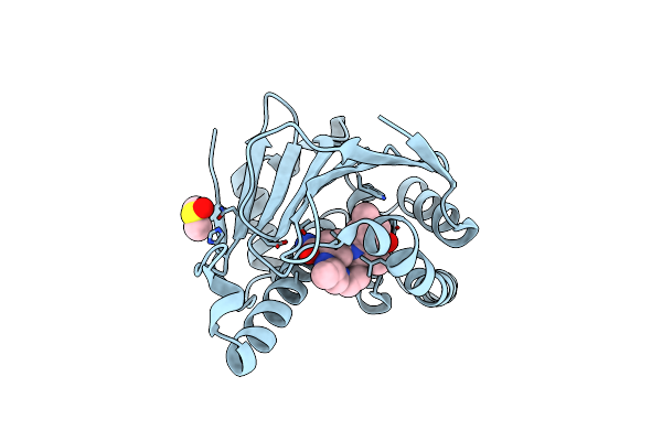
Organism: Homo sapiens
Method: X-RAY DIFFRACTION Resolution:2.20 Å Release Date: 2010-02-16 Classification: TRANSFERASE Ligands: 8I1 |
 |
Organism: Homo sapiens
Method: X-RAY DIFFRACTION Resolution:2.40 Å Release Date: 2010-02-09 Classification: TRANSFERASE Ligands: 8H1, SO4 |
 |
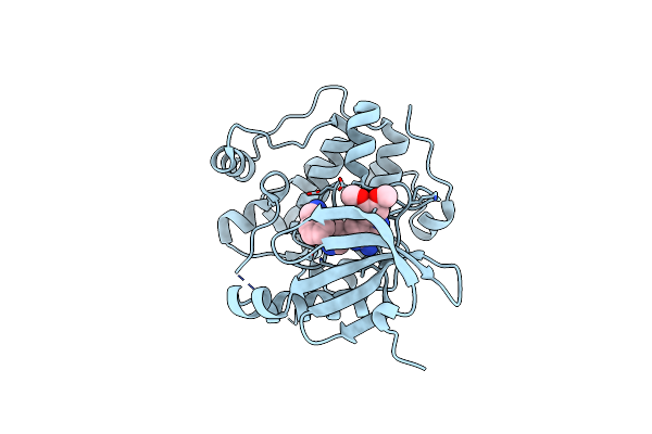
Phosphoinositide-Dependent Protein Kinase 1 (Pdk-1) In Complex With Compound 9
Organism: Homo sapiens
Method: X-RAY DIFFRACTION Resolution:2.30 Å Release Date: 2009-08-25 Classification: TRANSFERASE Ligands: 9BD |
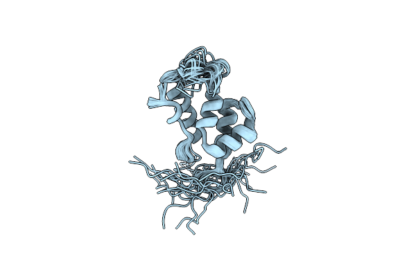 |
Solution Structure Of The W184A/M185A Mutant Of The Carboxy-Terminal Dimerization Domain Of The Hiv-1 Capsid Protein
Organism: Human immunodeficiency virus 1
Method: SOLUTION NMR Release Date: 2008-02-26 Classification: VIRAL PROTEIN |
 |
Solution Structure Of A Double Mutant Of The Carboxy-Terminal Dimerization Domain Of The Hiv-1 Capsid Protein
Organism: Human immunodeficiency virus 1
Method: SOLUTION NMR Release Date: 2008-02-12 Classification: VIRAL PROTEIN |
 |
Organism: African swine fever virus
Method: SOLUTION NMR Release Date: 2001-10-31 Classification: VIRAL PROTEIN |
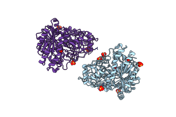 |
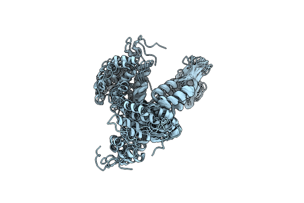
The Crystal Structure Of Formyltetrahydrofolate Synthetase From Moorella Thermoacetica
Organism: Moorella thermoacetica
Method: X-RAY DIFFRACTION Resolution:2.50 Å Release Date: 2001-02-14 Classification: LIGASE Ligands: SO4 |