
Search Count: 223
 |
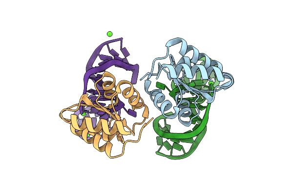
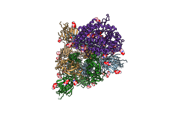
Cryo-Em Structure Of Chalcone Synthase (Chs) From Physcomitrella Patens In The Presence Of Chil
Organism: Physcomitrium patens
Method: ELECTRON MICROSCOPY Release Date: 2025-10-01 Classification: TRANSFERASE |
 |
Organism: Cytaeis uchidae
Method: X-RAY DIFFRACTION Resolution:1.65 Å Release Date: 2025-05-21 Classification: FLUORESCENT PROTEIN Ligands: CL, EDO |
 |
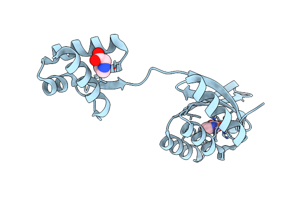
Crystal Structure Of The L7Ae Derivative Protein Ls4 In Complex With Its Co-Evolved Target Cs1 Rna
Organism: Archaeoglobus fulgidus (strain atcc 49558 / dsm 4304 / jcm 9628 / nbrc 100126 / vc-16), Synthetic construct
Method: X-RAY DIFFRACTION Resolution:1.89 Å Release Date: 2025-04-02 Classification: RNA BINDING PROTEIN Ligands: MG |
 |
Crystal Structure Of The L7Ae Derivative Protein Ls12 In Complex With Its Co-Evolved Target Cs2 Rna
Organism: Archaeoglobus fulgidus (strain atcc 49558 / dsm 4304 / jcm 9628 / nbrc 100126 / vc-16), Synthetic construct
Method: X-RAY DIFFRACTION Resolution:3.12 Å Release Date: 2025-04-02 Classification: RNA BINDING PROTEIN Ligands: CA |
 |
Organism: Archaeoglobus fulgidus dsm 4304
Method: X-RAY DIFFRACTION Resolution:1.80 Å Release Date: 2025-04-02 Classification: RNA BINDING PROTEIN |
 |
Organism: Thermotoga maritima
Method: X-RAY DIFFRACTION Resolution:2.30 Å Release Date: 2025-01-22 Classification: DNA BINDING PROTEIN Ligands: FE2, NIO, PRO |
 |
 |
Organism: Homo sapiens
Method: X-RAY DIFFRACTION Resolution:2.90 Å Release Date: 2024-11-06 Classification: IMMUNE SYSTEM |
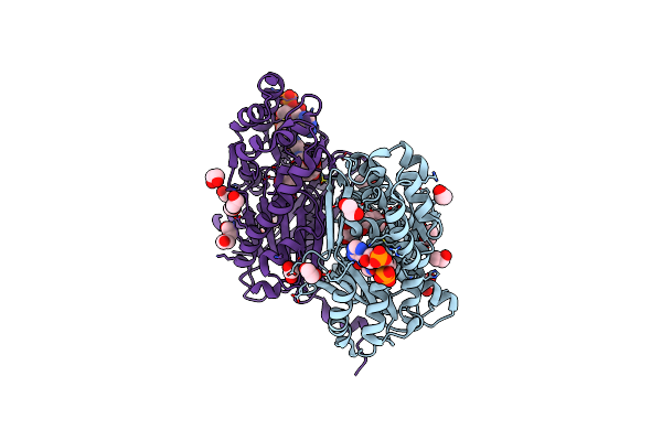 |
Chalcone Synthase From Glycine Max (L.) Merr (Soybean) Complexed With Naringenin And Coenzyme A
Organism: Glycine max
Method: X-RAY DIFFRACTION Resolution:1.83 Å Release Date: 2024-06-19 Classification: TRANSFERASE Ligands: COA, NAR, EDO, PGE, PEG |
 |
Organism: Microchaete diplosiphon
Method: X-RAY DIFFRACTION Resolution:1.75 Å Release Date: 2024-05-15 Classification: SIGNALING PROTEIN Ligands: CYC |
 |
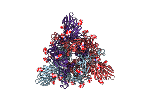
Structure Of The Sars-Cov-2 Eg.5.1 Spike Glycoprotein In Complex With Ace2 (1-Up State)
Organism: Severe acute respiratory syndrome coronavirus 2, Homo sapiens
Method: ELECTRON MICROSCOPY Release Date: 2024-05-01 Classification: VIRAL PROTEIN/PROTEIN BINDING Ligands: NAG |
 |
Organism: Homo sapiens, Severe acute respiratory syndrome coronavirus 2
Method: ELECTRON MICROSCOPY Release Date: 2024-05-01 Classification: VIRAL PROTEIN/PROTEIN BINDING Ligands: NAG |
 |
Organism: Severe acute respiratory syndrome coronavirus 2
Method: ELECTRON MICROSCOPY Release Date: 2024-04-24 Classification: VIRAL PROTEIN Ligands: NAG |
 |
Organism: Severe acute respiratory syndrome coronavirus 2
Method: ELECTRON MICROSCOPY Release Date: 2024-04-24 Classification: VIRAL PROTEIN Ligands: NAG |
 |
Organism: Homo sapiens
Method: ELECTRON MICROSCOPY Resolution:3.50 Å Release Date: 2024-03-06 Classification: CONTRACTILE PROTEIN Ligands: ADP, MG |
 |
Neutron Structure Of Bacillus Thermoproteolyticus Ferredoxin At Room Temperature
Organism: Bacillus thermoproteolyticus
Method: X-RAY DIFFRACTION, NEUTRON DIFFRACTION Resolution:1.45 Å, 1.60 Å Release Date: 2024-02-07 Classification: ELECTRON TRANSPORT Ligands: SF4 |
 |
Organism: Cytaeis uchidae
Method: X-RAY DIFFRACTION Resolution:1.60 Å Release Date: 2023-07-19 Classification: FLUORESCENT PROTEIN Ligands: CL, EDO |
 |
Organism: Severe acute respiratory syndrome coronavirus 2
Method: ELECTRON MICROSCOPY Release Date: 2023-05-24 Classification: VIRAL PROTEIN Ligands: NAG |
 |
Organism: Severe acute respiratory syndrome coronavirus 2
Method: ELECTRON MICROSCOPY Release Date: 2023-05-24 Classification: VIRAL PROTEIN Ligands: NAG |
 |
Structure Of Sars-Cov-2 Xbb.1 Spike Glycoprotein In Complex With Ace2 (1-Up State)
Organism: Severe acute respiratory syndrome coronavirus 2, Homo sapiens
Method: ELECTRON MICROSCOPY Release Date: 2023-05-24 Classification: VIRAL PROTEIN/PROTEIN BINDING Ligands: NAG |