
Search Count: 115
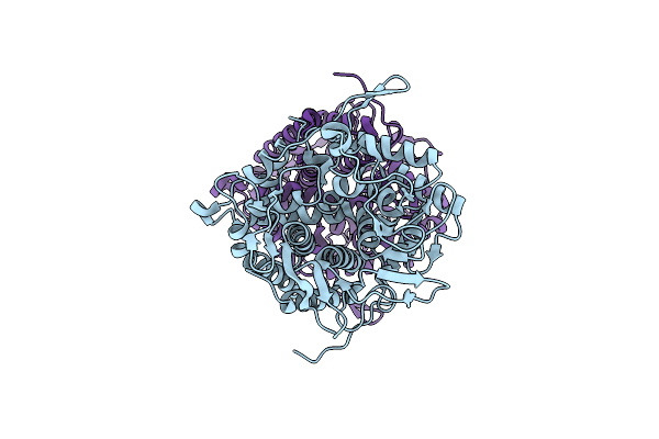 |
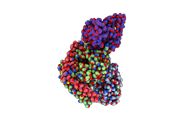
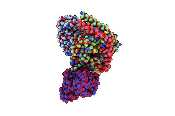
Structure Of Beta-1,2-Glucanase From Endozoicomonas Elysicola (Eesgl1, Ligand-Free)
Organism: Endozoicomonas elysicola dsm 22380
Method: X-RAY DIFFRACTION Release Date: 2025-01-22 Classification: HYDROLASE |
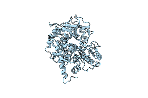 |
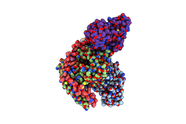
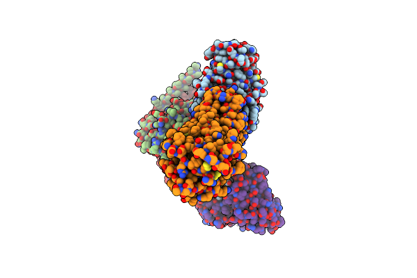
Structure Of Beta-1,2-Glucanase From Photobacterium Gaetbulicola (Pgsgl3, Ligand-Free)
Organism: Photobacterium gaetbulicola gung47
Method: X-RAY DIFFRACTION Release Date: 2025-01-22 Classification: HYDROLASE Ligands: CL |
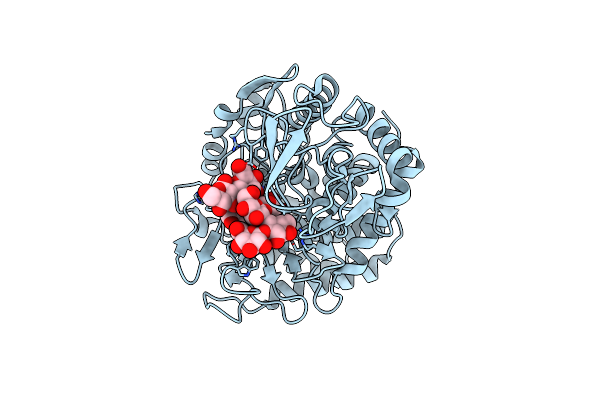 |
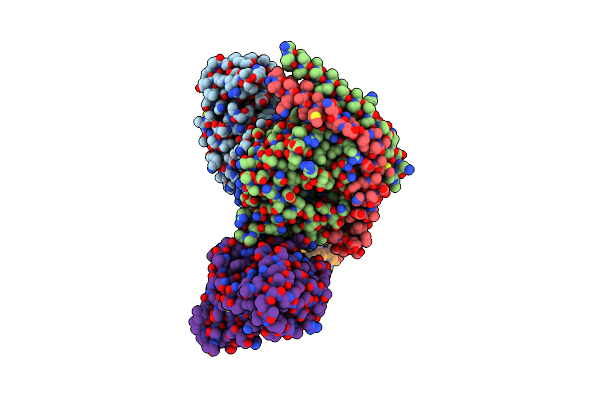
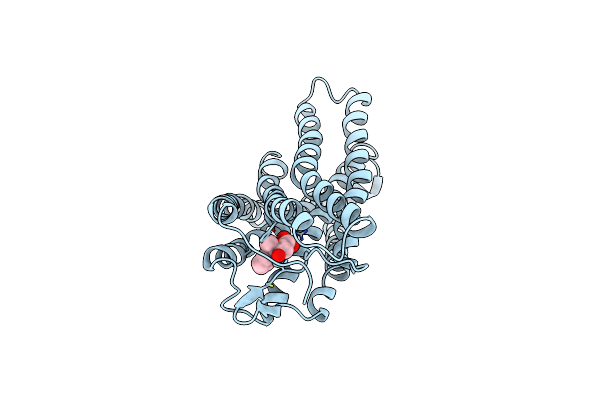
Structure Of Beta-1,2-Glucanase From Xanthomonas Campestris Pv. Campestris (Beta-1,2-Glucoheptasaccharide Complex)-E239Q Mutant
Organism: Xanthomonas campestris pv. campestris
Method: X-RAY DIFFRACTION Release Date: 2025-01-22 Classification: HYDROLASE |
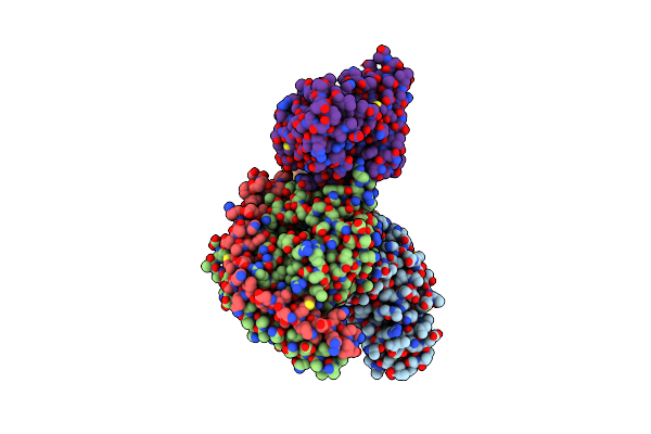 |
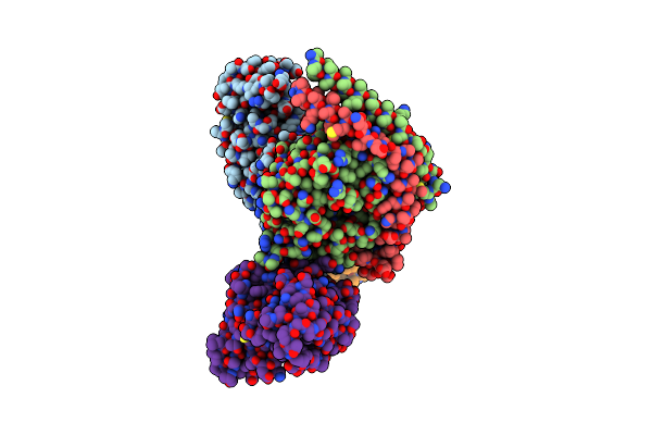
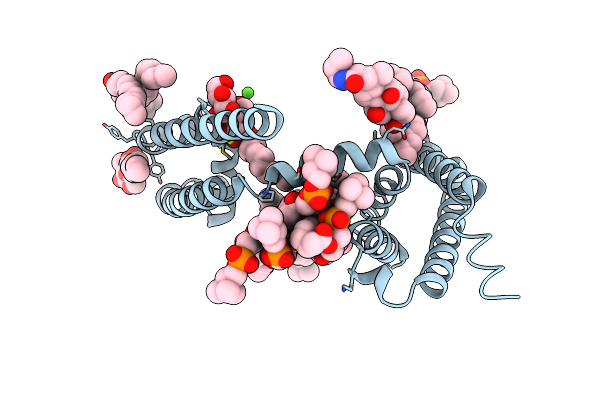
Organism: Escherichia coli, Homo sapiens, Synthetic construct, Mus musculus
Method: ELECTRON MICROSCOPY Release Date: 2025-01-01 Classification: MEMBRANE PROTEIN Ligands: NKP |
 |
Organism: Homo sapiens, Mus musculus, Escherichia coli, Synthetic construct
Method: ELECTRON MICROSCOPY Release Date: 2025-01-01 Classification: MEMBRANE PROTEIN Ligands: NKP |
 |
Organism: Escherichia coli, Homo sapiens, Synthetic construct, Mus musculus
Method: ELECTRON MICROSCOPY Release Date: 2025-01-01 Classification: MEMBRANE PROTEIN Ligands: NKP |
 |
Organism: Mus musculus
Method: ELECTRON MICROSCOPY Resolution:2.70 Å Release Date: 2024-07-31 Classification: HYDROLASE Ligands: A1L0C |
 |
Organism: Canis lupus familiaris
Method: ELECTRON MICROSCOPY Resolution:2.20 Å Release Date: 2024-07-31 Classification: HYDROLASE Ligands: A1L0D |
 |
Organism: Canis lupus familiaris
Method: ELECTRON MICROSCOPY Resolution:2.00 Å Release Date: 2024-07-31 Classification: HYDROLASE Ligands: A1L0C |
 |
Organism: Mus musculus
Method: ELECTRON MICROSCOPY Resolution:2.50 Å Release Date: 2024-07-31 Classification: HYDROLASE Ligands: A1L0D, THR |
 |
Organism: Escherichia coli, Homo sapiens, Synthetic construct, Mus musculus
Method: ELECTRON MICROSCOPY Release Date: 2023-09-13 Classification: SIGNALING PROTEIN Ligands: NAG, OKL |
 |
Organism: Homo sapiens, Synthetic construct, Mus musculus, Escherichia coli
Method: ELECTRON MICROSCOPY Release Date: 2023-08-30 Classification: SIGNALING PROTEIN Ligands: NAG, FI7 |
 |
Organism: Escherichia coli, Homo sapiens, Mus musculus, Synthetic construct
Method: ELECTRON MICROSCOPY Release Date: 2023-08-30 Classification: SIGNALING PROTEIN Ligands: NAG, P8A |
 |
Organism: Homo sapiens, Synthetic construct, Mus musculus, Escherichia coli
Method: ELECTRON MICROSCOPY Release Date: 2023-08-30 Classification: SIGNALING PROTEIN Ligands: NAG, P9X |
 |
Organism: Escherichia coli, Homo sapiens, Synthetic construct, Mus musculus
Method: ELECTRON MICROSCOPY Release Date: 2023-08-30 Classification: SIGNALING PROTEIN Ligands: P9X |
 |
Organism: Escherichia coli, Homo sapiens
Method: ELECTRON MICROSCOPY Release Date: 2023-08-30 Classification: SIGNALING PROTEIN Ligands: P9X |
 |
Crystal Structure Of Voltage-Gated Sodium Channel Navab N49K Mutant In Sodium Ion Condition
Organism: Aliarcobacter butzleri
Method: X-RAY DIFFRACTION Resolution:3.30 Å Release Date: 2023-07-26 Classification: MEMBRANE PROTEIN Ligands: LMT, 1N7, PX4, NA |
 |
Crystal Structure Of Voltage-Gated Sodium Channel Navab N49K Mutant In Calcium Ion Condition
Organism: Aliarcobacter butzleri
Method: X-RAY DIFFRACTION Resolution:2.70 Å Release Date: 2023-07-26 Classification: MEMBRANE PROTEIN Ligands: LMT, 1N7, PX4, CA |
 |
Crystal Structure Of Voltage-Gated Sodium Channel Navab N49K/L176Q Mutant In Sodium Ion Condition
Organism: Aliarcobacter butzleri
Method: X-RAY DIFFRACTION Resolution:3.40 Å Release Date: 2023-07-26 Classification: MEMBRANE PROTEIN Ligands: LMT, 1N7, PX4, NA |
 |
Crystal Structure Of Voltage-Gated Sodium Channel Navab N49K/L176Q Mutant In Calcium Ion Condition
Organism: Aliarcobacter butzleri
Method: X-RAY DIFFRACTION Resolution:3.40 Å Release Date: 2023-07-26 Classification: MEMBRANE PROTEIN Ligands: LMT, 1N7, PX4, CA |