
Search Count: 307
 |
Organism: Mirabilis jalapa
Method: X-RAY DIFFRACTION Release Date: 2025-12-03 Classification: ANTIVIRAL PROTEIN Ligands: SO4 |
 |
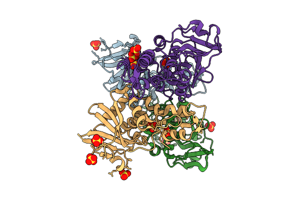
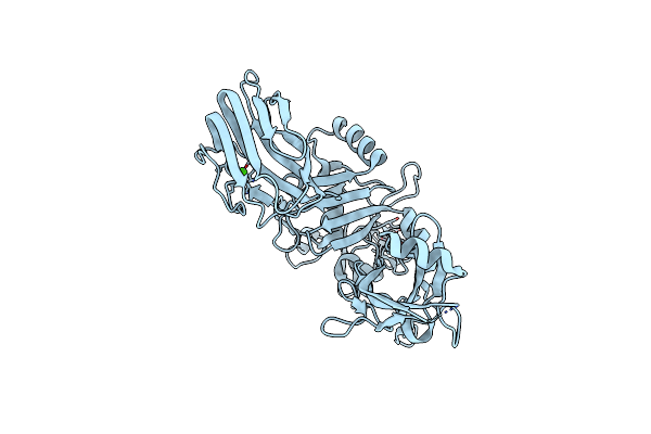
Organism: Homo sapiens, Bos taurus
Method: X-RAY DIFFRACTION Release Date: 2025-06-11 Classification: TRANSFERASE/INHIBITOR Ligands: A1L62 |
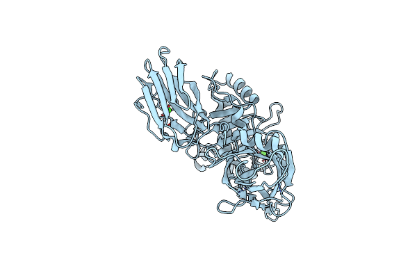 |
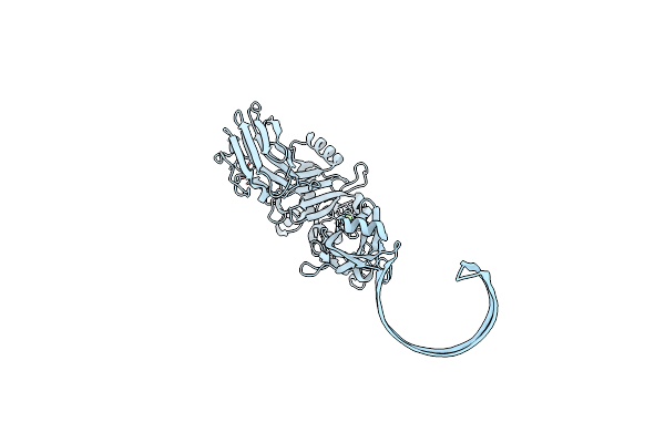
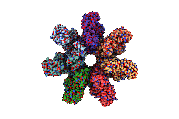
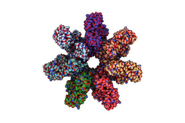
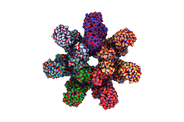
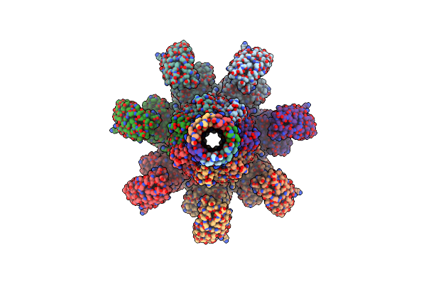
Organism: Clostridium perfringens
Method: ELECTRON MICROSCOPY Release Date: 2025-05-14 Classification: TOXIN Ligands: CA |
 |
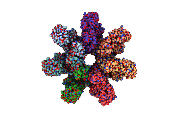
Organism: Clostridium perfringens
Method: ELECTRON MICROSCOPY Release Date: 2025-05-14 Classification: TOXIN Ligands: CA |
 |
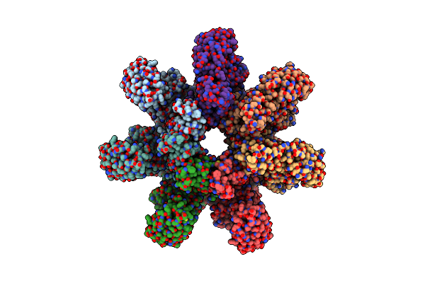
Organism: Clostridium perfringens
Method: ELECTRON MICROSCOPY Release Date: 2025-05-14 Classification: TOXIN Ligands: CA |
 |
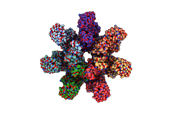
Organism: Clostridium perfringens
Method: ELECTRON MICROSCOPY Release Date: 2025-05-14 Classification: TOXIN Ligands: CA |
 |
Organism: Clostridium perfringens
Method: ELECTRON MICROSCOPY Release Date: 2025-05-14 Classification: TOXIN Ligands: CA |
 |
Organism: Clostridium perfringens
Method: ELECTRON MICROSCOPY Release Date: 2025-05-14 Classification: TOXIN Ligands: CA |
 |
Organism: Clostridium perfringens
Method: ELECTRON MICROSCOPY Release Date: 2025-05-14 Classification: TOXIN Ligands: CA |
 |
Organism: Clostridium perfringens
Method: ELECTRON MICROSCOPY Release Date: 2025-05-14 Classification: TOXIN Ligands: CA |
 |
Organism: Clostridium perfringens
Method: ELECTRON MICROSCOPY Release Date: 2025-05-14 Classification: TOXIN Ligands: CA |
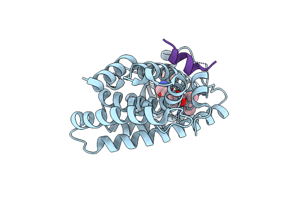 |
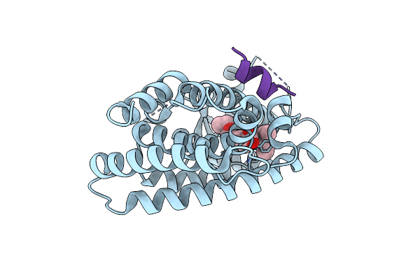
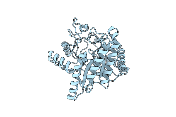
Human Ppar Alpha Ligand Binding Domain In Complex With A 1H-Pyrazolo[3,4-B]Pyridine-Derived Compound
Organism: Homo sapiens
Method: X-RAY DIFFRACTION Resolution:1.77 Å Release Date: 2025-04-09 Classification: TRANSCRIPTION Ligands: A1LZ9 |
 |
Human Ppar Alpha Ligand Binding Domain In Complex With A 1H-Pyrazolo[3,4-B]Pyridine-Derived Compound
Organism: Homo sapiens
Method: X-RAY DIFFRACTION Resolution:2.01 Å Release Date: 2025-04-09 Classification: TRANSCRIPTION Ligands: A1L0A |
 |
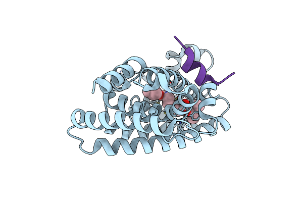
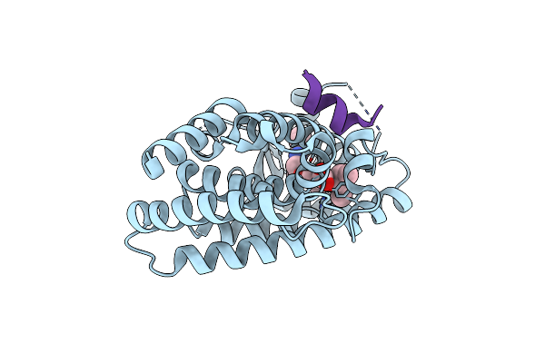
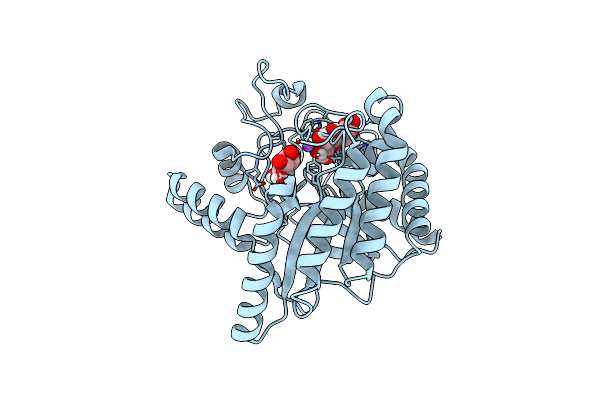
Human Ppar Alpha Ligand Binding Domain In Complex With A 1H-Pyrazolo[3,4-B]Pyridine-Derived Compound
Organism: Homo sapiens
Method: X-RAY DIFFRACTION Resolution:1.85 Å Release Date: 2025-04-02 Classification: TRANSCRIPTION Ligands: A1LZX |
 |
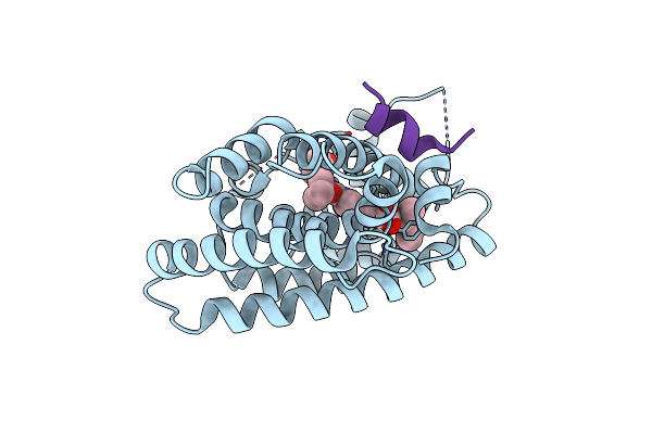
Human Ppar Alpha Ligand Binding Domain In Complex With A 1H-Pyrazolo[3,4-B]Pyridine-Derived Compound
Organism: Homo sapiens
Method: X-RAY DIFFRACTION Resolution:1.59 Å Release Date: 2025-04-02 Classification: TRANSCRIPTION Ligands: A1LZY |
 |
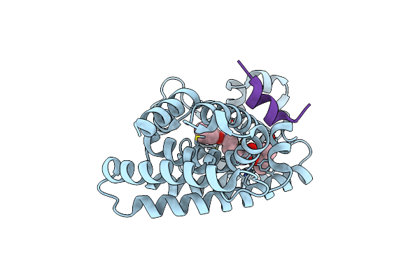
Human Ppar Alpha Ligand Binding Domain In Complex With A 1H-Pyrazolo[3,4-B]Pyridine-Derived Compound
Organism: Homo sapiens
Method: X-RAY DIFFRACTION Resolution:2.00 Å Release Date: 2025-04-02 Classification: TRANSCRIPTION Ligands: PGO, A1LZ0 |
 |
Human Ppar Alpha Ligand Binding Domain In Complex With A 1H-Pyrazolo[3,4-B]Pyridine-Derived Compound
Organism: Homo sapiens
Method: X-RAY DIFFRACTION Resolution:1.95 Å Release Date: 2025-04-02 Classification: TRANSCRIPTION Ligands: A1LZ1 |
 |
Organism: Clostridium perfringens
Method: ELECTRON MICROSCOPY Release Date: 2025-03-26 Classification: TOXIN Ligands: CA |
 |
Neutron Structure Of Cellulase Cel6A From Phanerochaete Chrysosporium At Room Temperature
Organism: Phanerodontia chrysosporium
Method: X-RAY DIFFRACTION, NEUTRON DIFFRACTION Resolution:1.36 Å, 1.86 Å Release Date: 2025-03-12 Classification: HYDROLASE |
 |
Neutron Structure Of Cellulase Cel6A From Phanerochaete Chrysosporium At Room Temperature, Enzyme-Product Complex
Organism: Phanerodontia chrysosporium
Method: X-RAY DIFFRACTION, NEUTRON DIFFRACTION Resolution:1.40 Å, 1.8 Å Release Date: 2025-03-12 Classification: HYDROLASE Ligands: BGC, NA |