Search Count: 918
 |
Organism: Homo sapiens
Method: ELECTRON MICROSCOPY Release Date: 2025-07-23 Classification: OXYGEN BINDING Ligands: HEM, CMO |
 |
Organism: Homo sapiens
Method: ELECTRON MICROSCOPY Release Date: 2025-07-23 Classification: OXYGEN BINDING Ligands: HEM, CMO |
 |
Organism: Alligator mississippiensis
Method: ELECTRON MICROSCOPY Release Date: 2025-07-23 Classification: OXYGEN BINDING Ligands: HEM, CMO |
 |
Organism: Alligator mississippiensis
Method: ELECTRON MICROSCOPY Release Date: 2025-07-23 Classification: OXYGEN BINDING Ligands: HEM, CMO |
 |
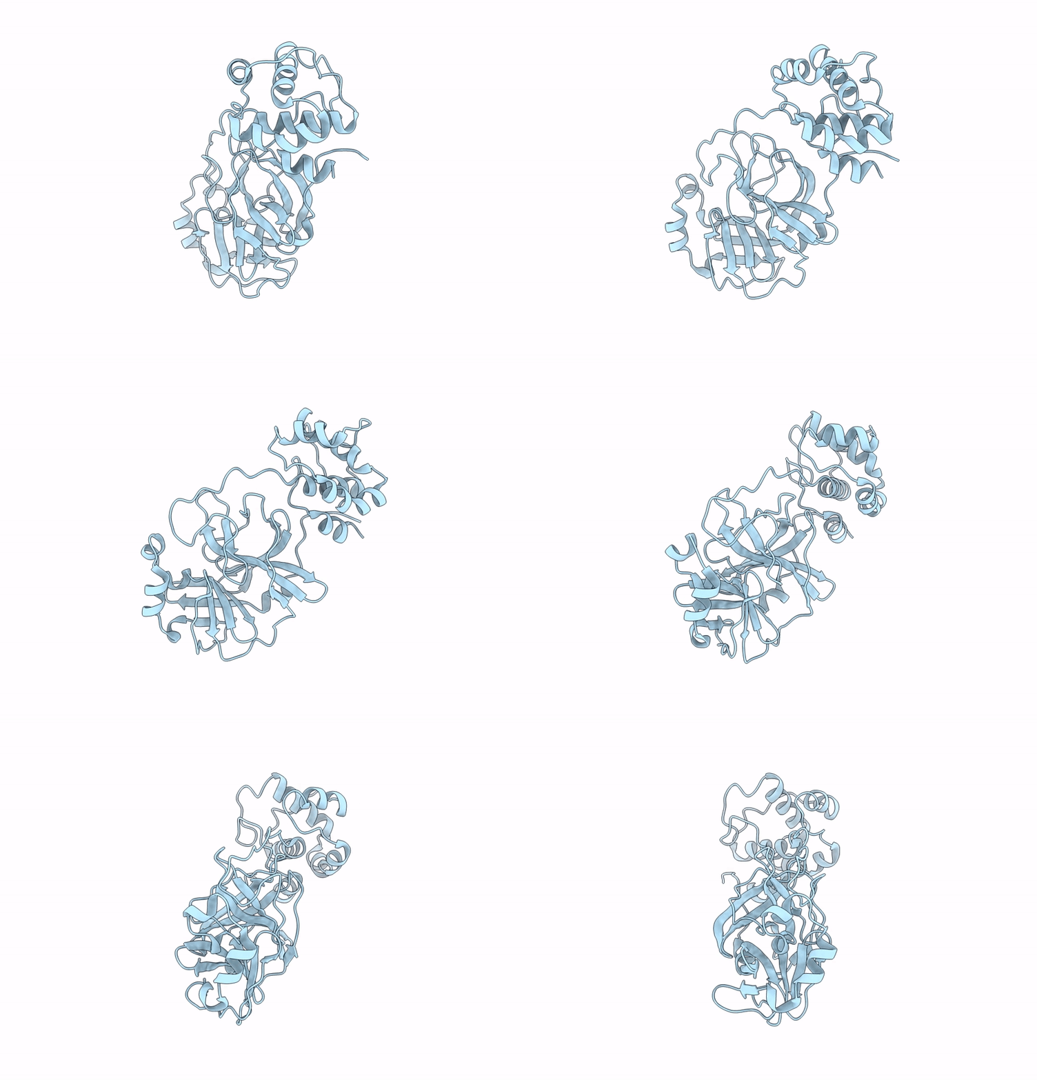
Organism: Homo sapiens
Method: X-RAY DIFFRACTION Release Date: 2025-07-16 Classification: TRANSPORT PROTEIN Ligands: A1L9H, PO4 |
 |
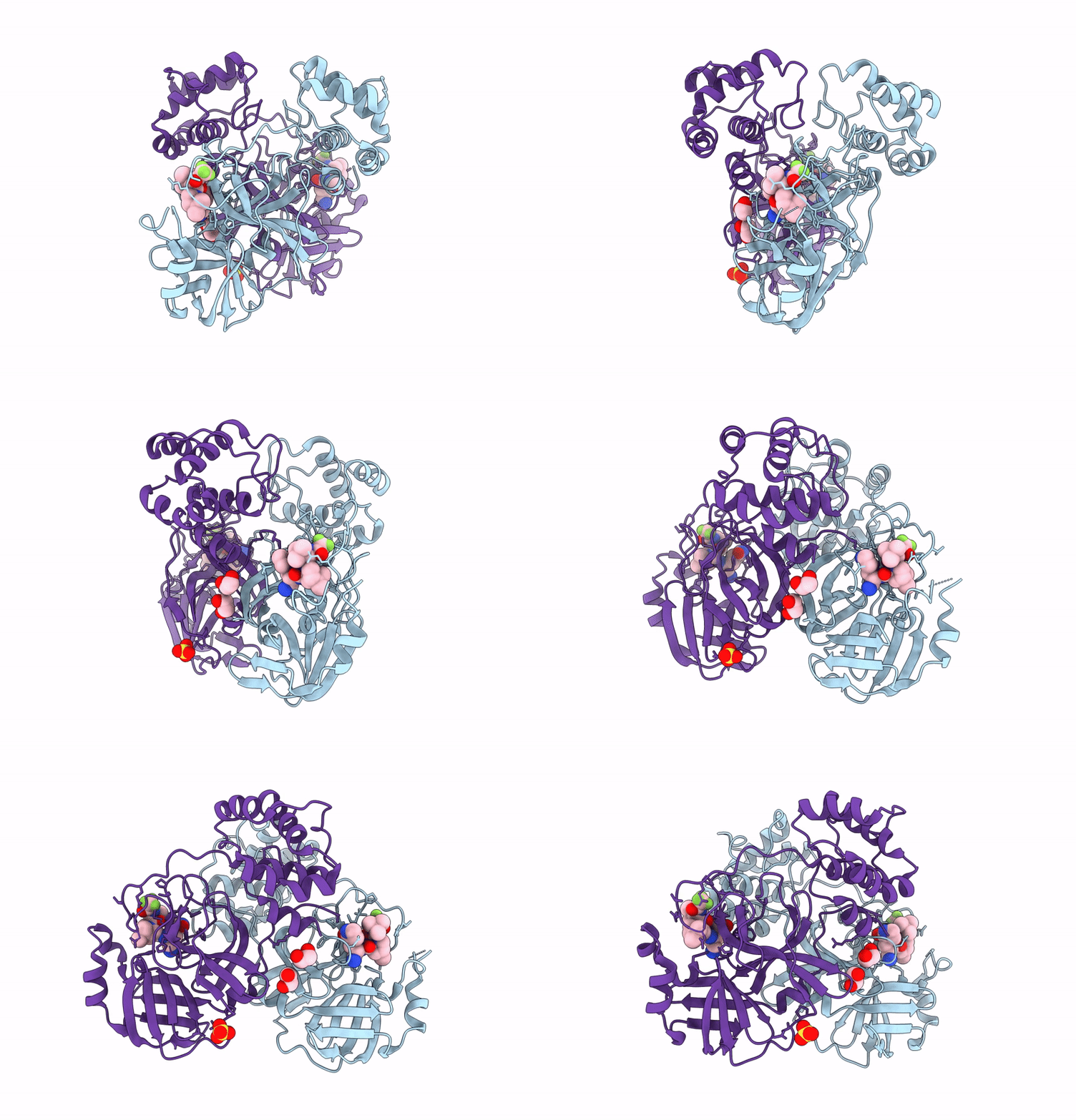
Organism: Severe acute respiratory syndrome coronavirus 2
Method: X-RAY DIFFRACTION Release Date: 2025-07-09 Classification: HYDROLASE |
 |
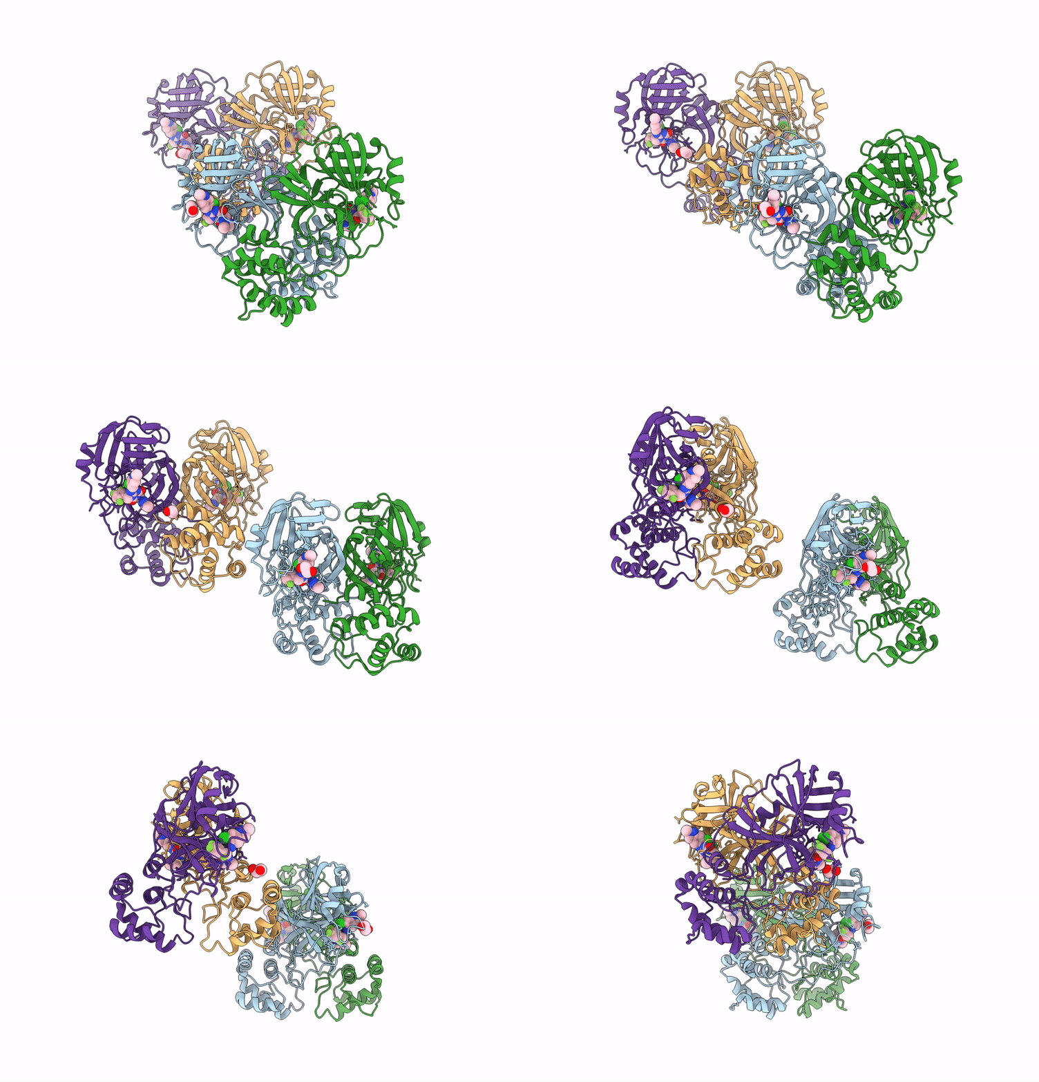
X-Ray Crystal Structure Of Sars-Cov-2 Main Protease Quadruple Mutants In Complex With Nirmatrelvir
Organism: Severe acute respiratory syndrome coronavirus 2
Method: X-RAY DIFFRACTION Release Date: 2025-07-09 Classification: HYDROLASE/INHIBITOR Ligands: 4WI, EDO, BR, SO4 |
 |
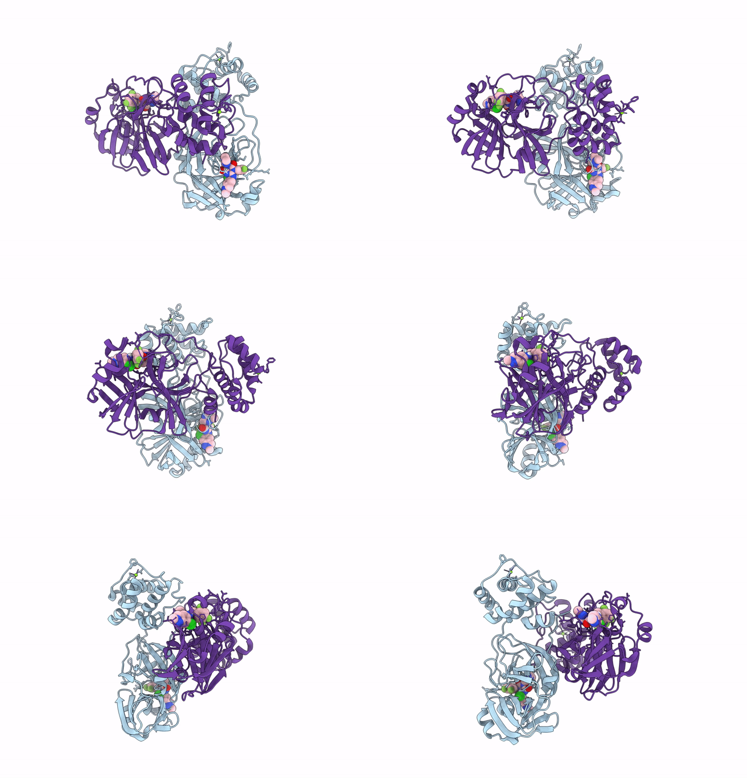
X-Ray Crystal Structure Of Sars-Cov-2 Main Protease Quadruple Mutants In Complex With Ensitrelvir
Organism: Severe acute respiratory syndrome coronavirus 2
Method: X-RAY DIFFRACTION Release Date: 2025-07-09 Classification: HYDROLASE/INHIBITOR Ligands: 7YY, EDO |
 |
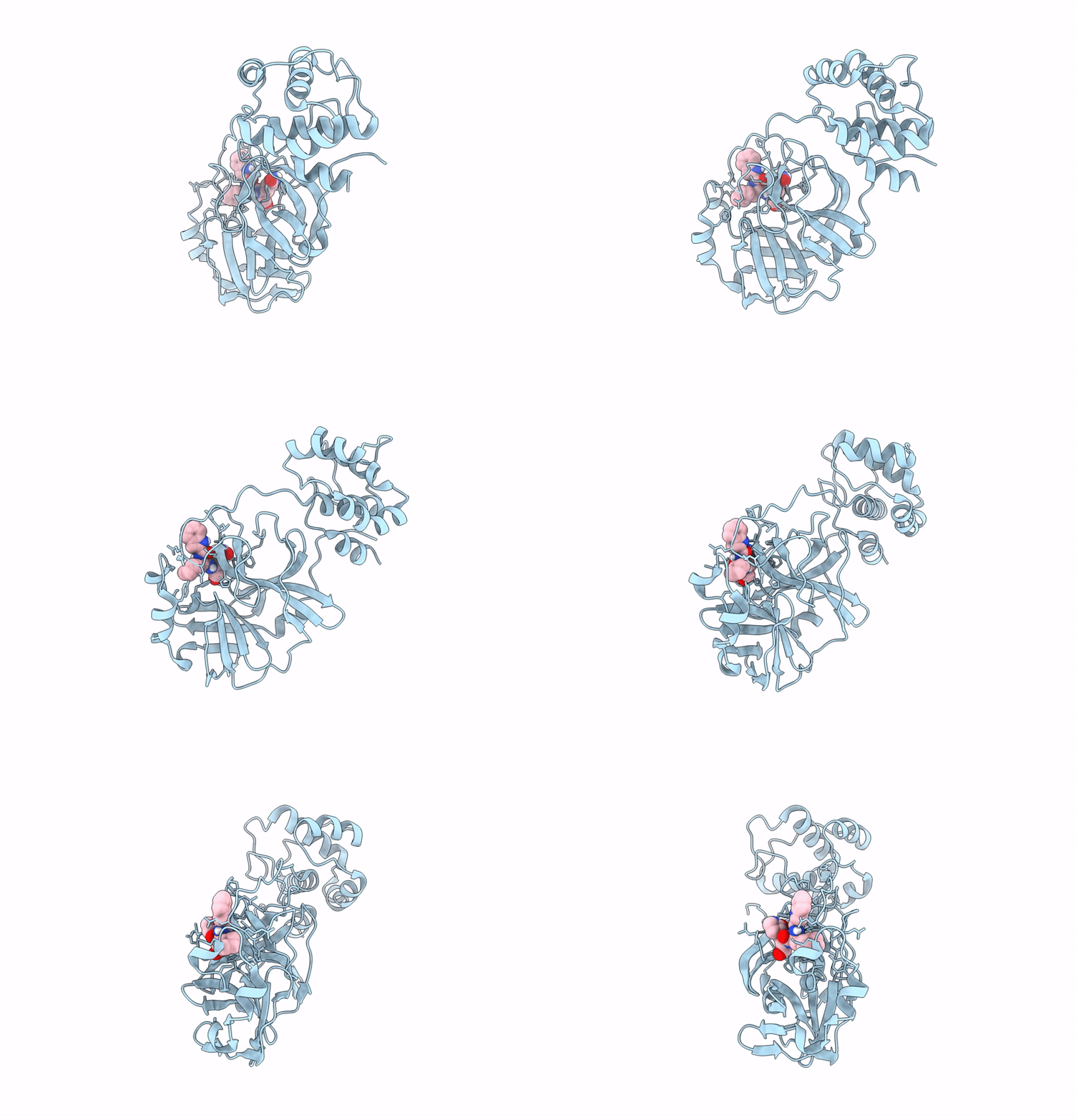
X-Ray Crystal Structure Of Sars-Cov-2 Main Protease Double Mutants In Complex With Ensitrelvir
Organism: Severe acute respiratory syndrome coronavirus 2
Method: X-RAY DIFFRACTION Release Date: 2025-07-09 Classification: HYDROLASE/INHIBITOR Ligands: 7YY, MG, CL |
 |
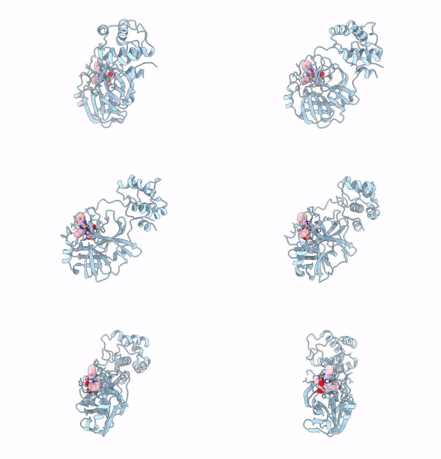
X-Ray Crystal Structure Of Sars-Cov-2 Main Protease Triple Mutants In Complex With Bofutrelvir
Organism: Severe acute respiratory syndrome coronavirus 2
Method: X-RAY DIFFRACTION Release Date: 2025-07-09 Classification: HYDROLASE/INHIBITOR Ligands: FHR |
 |
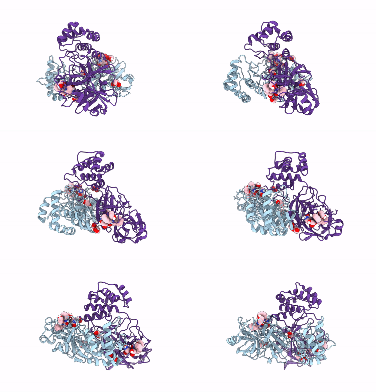
X-Ray Crystal Structure Of Sars-Cov-2 Main Protease Quadruple Mutants In Complex With Bofutrelvir
Organism: Severe acute respiratory syndrome coronavirus 2
Method: X-RAY DIFFRACTION Release Date: 2025-07-09 Classification: HYDROLASE/INHIBITOR Ligands: FHR |
 |
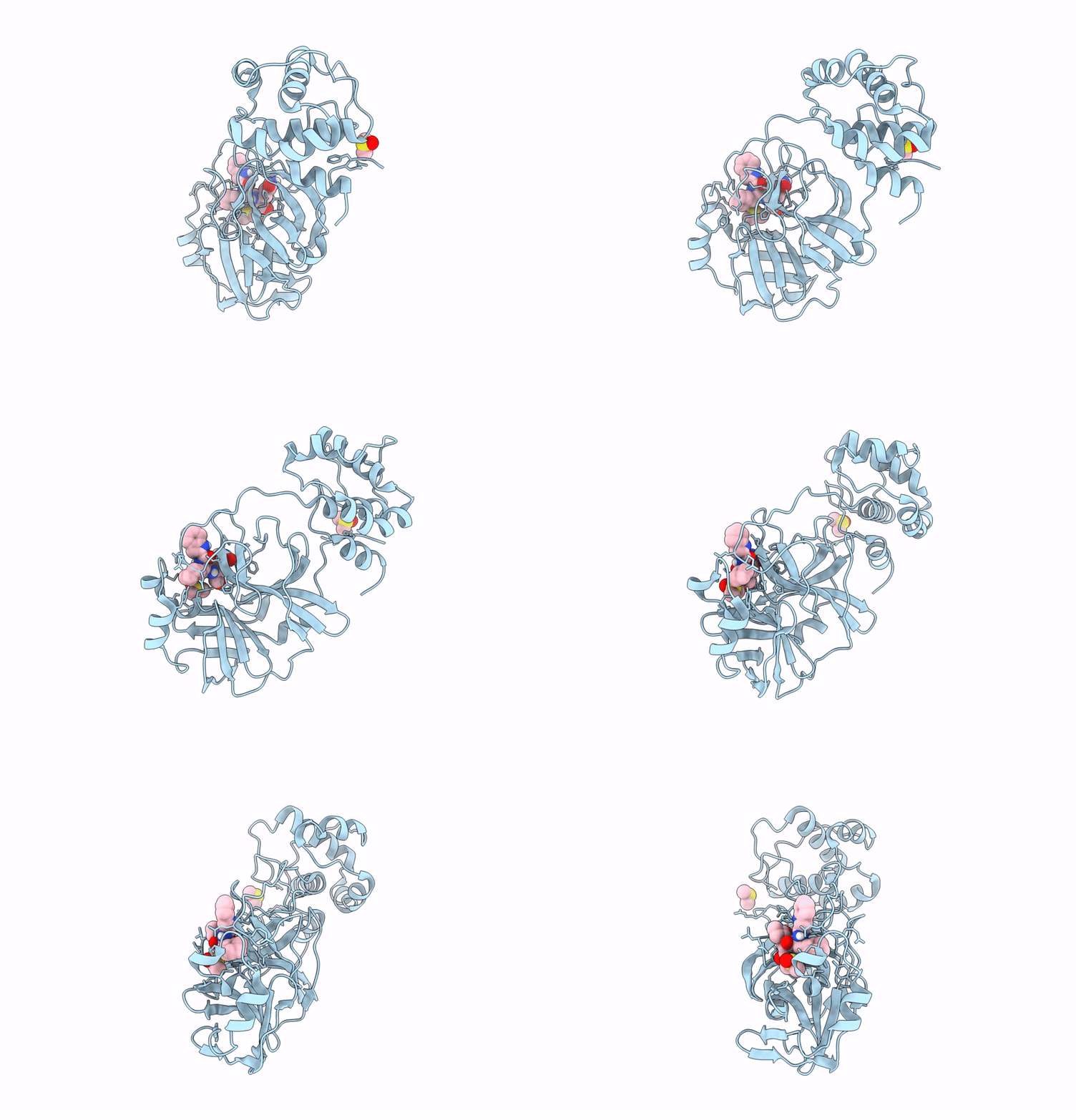
Structure Of Sars-Cov-2 Main Protease In Complex With Bofutrelvir In Orthorhombic Form
Organism: Severe acute respiratory syndrome coronavirus 2
Method: X-RAY DIFFRACTION Release Date: 2025-07-09 Classification: HYDROLASE/INHIBITOR Ligands: FHR, EDO, PEG |
 |
X-Ray Crystal Structure Of Sars-Cov-2 Main Protease Complex With Bofutrelvir
Organism: Severe acute respiratory syndrome coronavirus 2
Method: X-RAY DIFFRACTION Release Date: 2025-07-09 Classification: HYDROLASE/INHIBITOR Ligands: FHR, DMS |
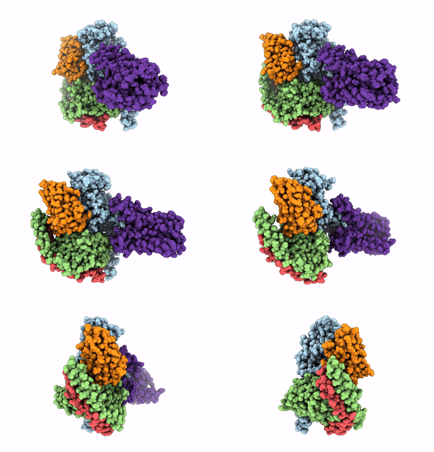 |
Organism: Homo sapiens, Rattus norvegicus, Bos taurus, Unidentified
Method: ELECTRON MICROSCOPY Release Date: 2025-06-11 Classification: MEMBRANE PROTEIN Ligands: HSM |
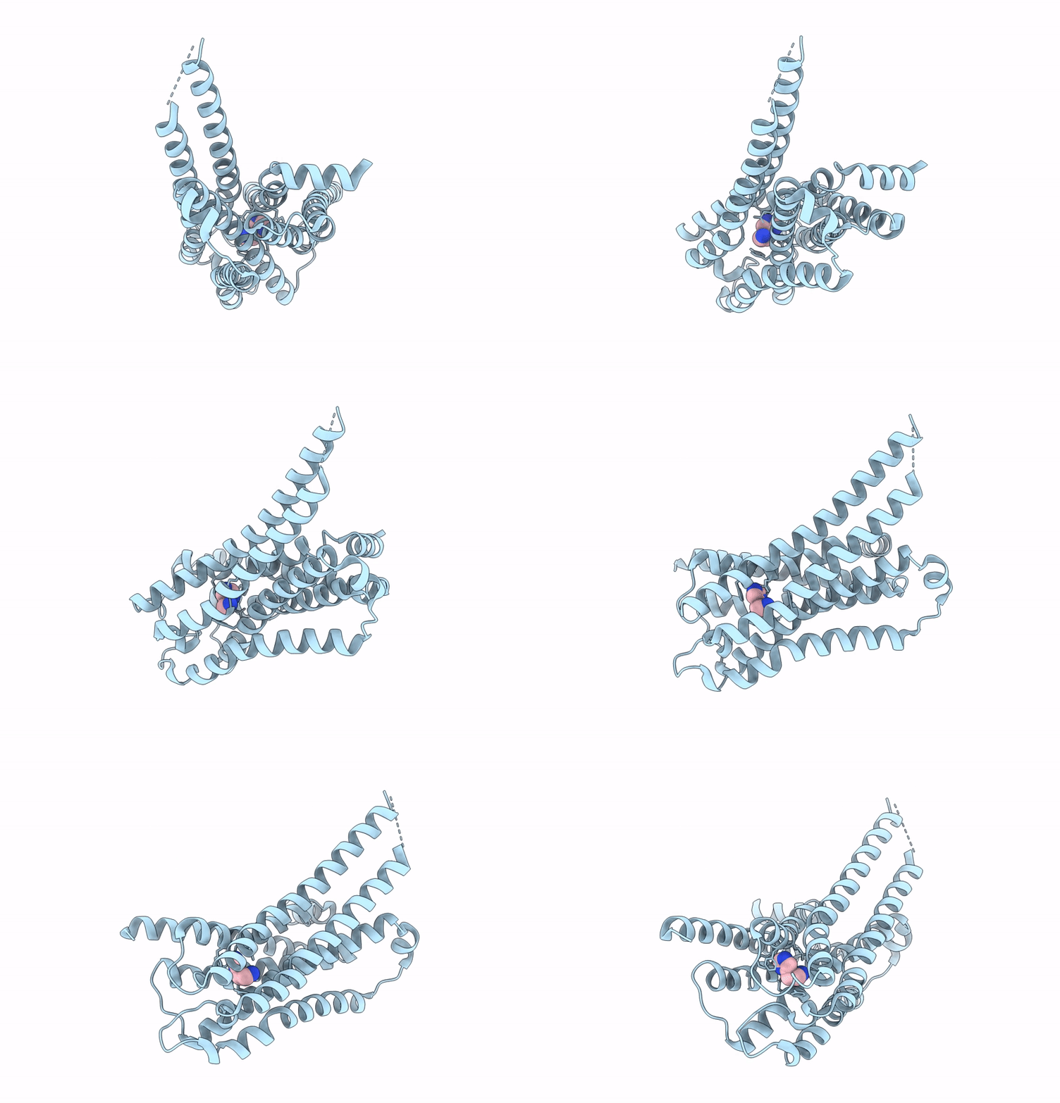 |
Cryo-Em Structure Of The Histamine H4 Receptor-Gi Protein Complex (Receptor Focused)
Organism: Homo sapiens
Method: ELECTRON MICROSCOPY Release Date: 2025-06-11 Classification: MEMBRANE PROTEIN Ligands: HSM |
 |
Organism: Homo sapiens, Rattus norvegicus, Bos taurus, Mus musculus
Method: ELECTRON MICROSCOPY Release Date: 2025-06-11 Classification: MEMBRANE PROTEIN Ligands: HSM |
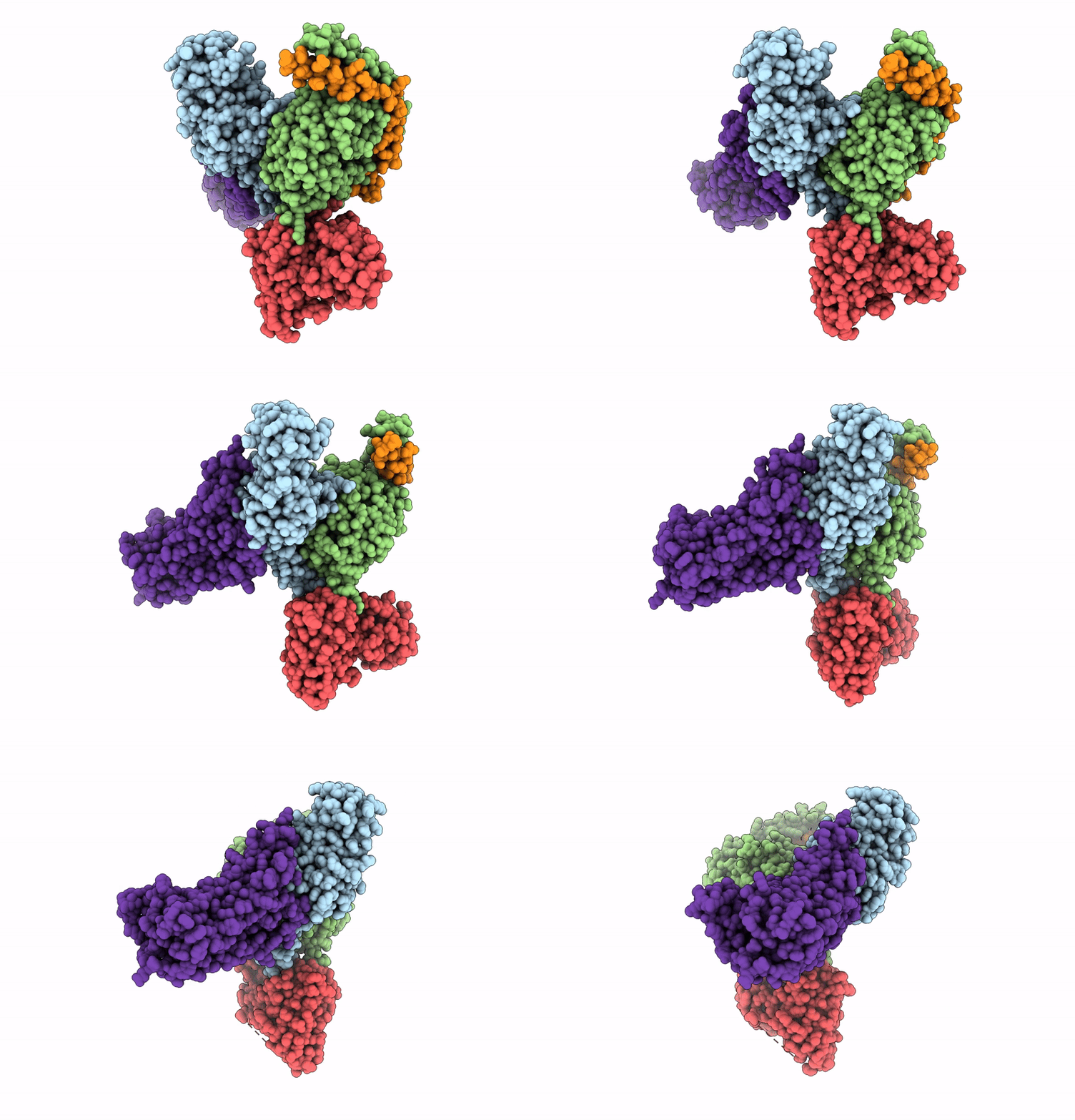 |
Cryo-Em Structure Of The Histamine H4 Receptor-Gi Protein Complex (Overall)
Organism: Homo sapiens, Rattus norvegicus, Mus musculus, Bos taurus
Method: ELECTRON MICROSCOPY Release Date: 2025-06-11 Classification: MEMBRANE PROTEIN Ligands: HSM |
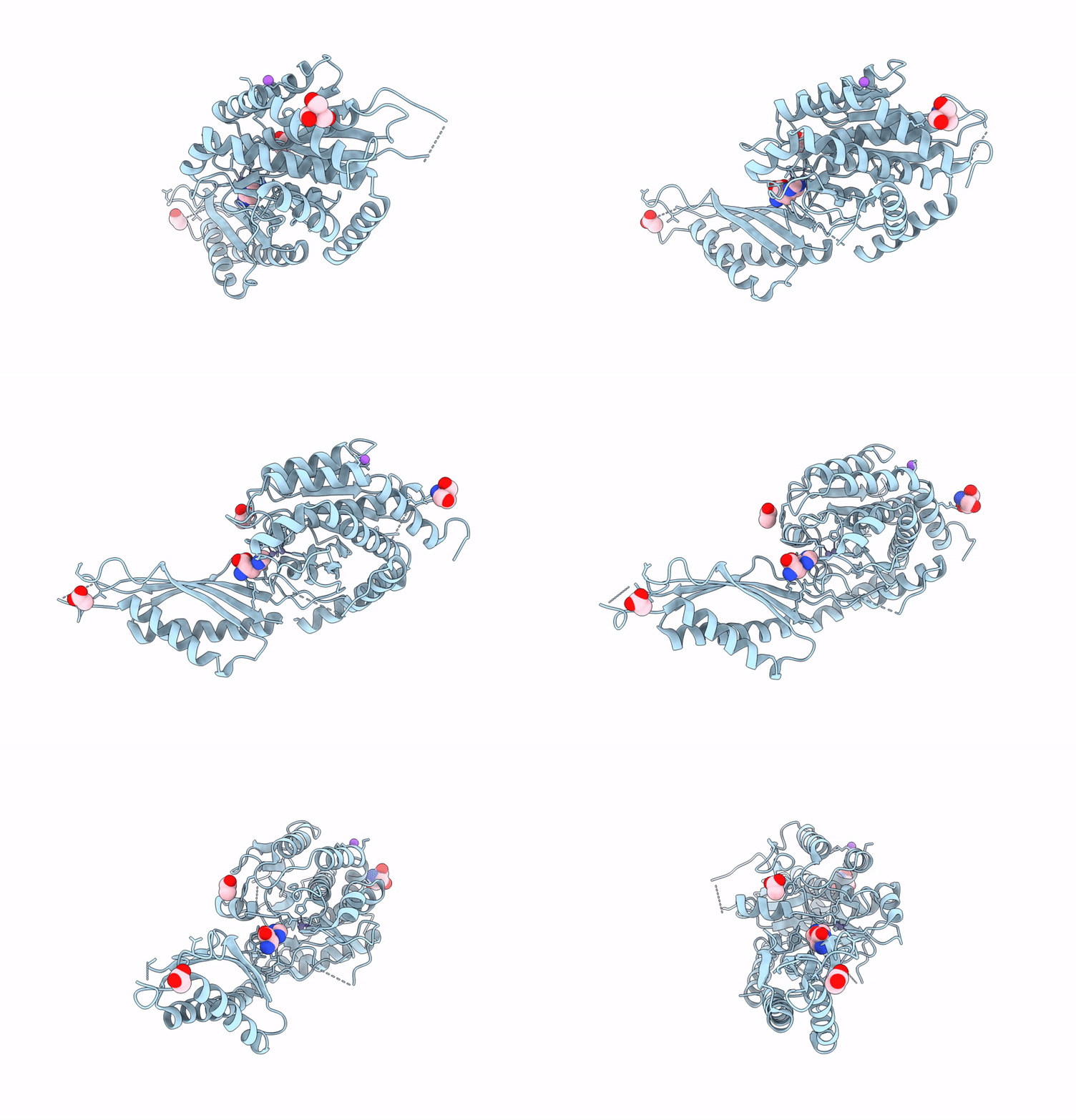 |
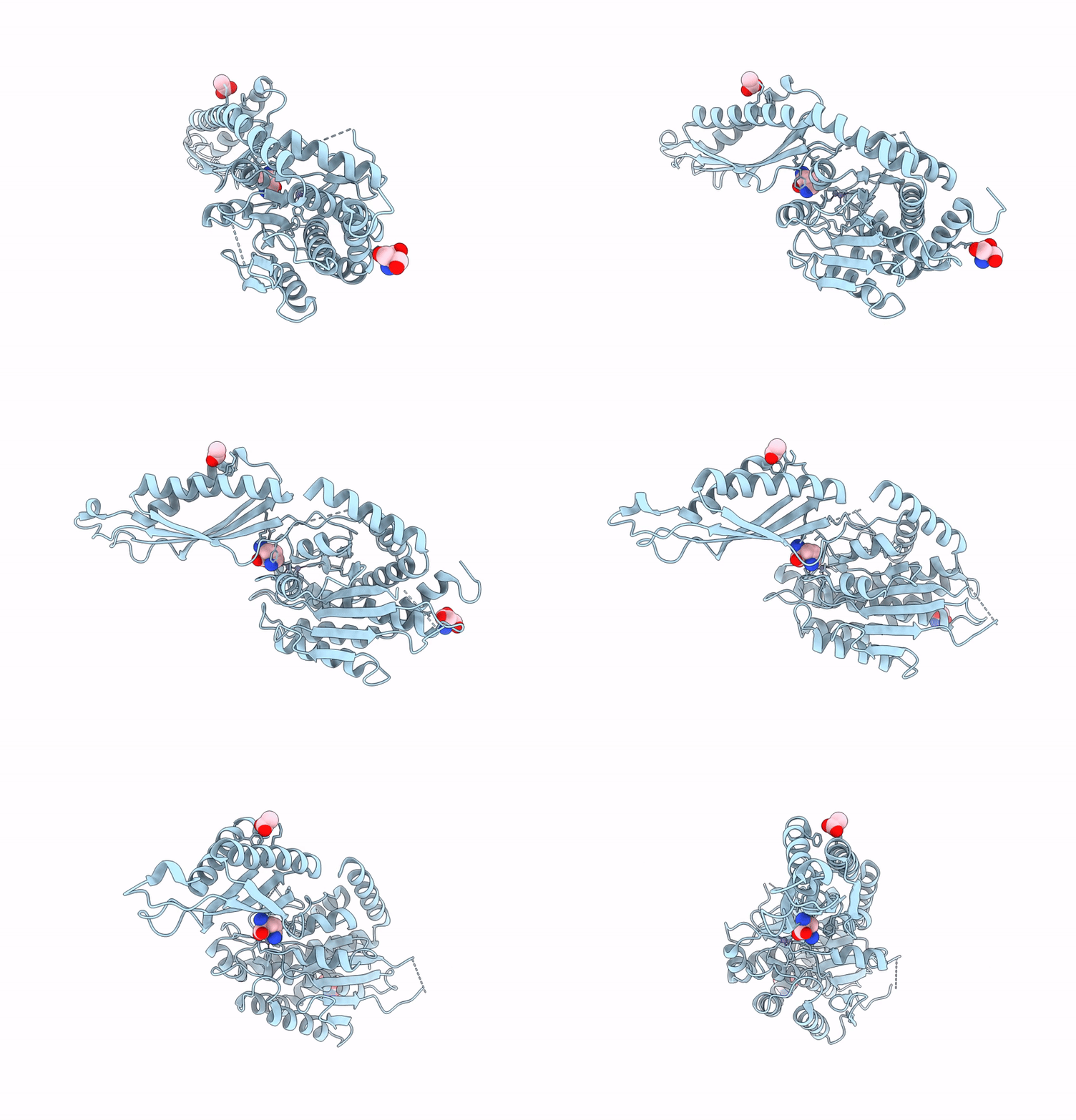
Crystal Structure Of Human Cytosolic Beta-Alanyl Lysine Dipeptidase With Crystal Soaked In Beta-Alanyl Histidine
Organism: Homo sapiens
Method: X-RAY DIFFRACTION Resolution:2.30 Å Release Date: 2025-05-14 Classification: HYDROLASE Ligands: ZN, HIS, TRS, ACY, NA |
 |
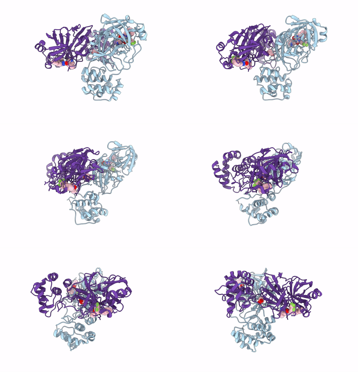
Crystal Structure Of Human Cytosolic Beta-Alanyl Lysine Dipeptidase Tyr314Phe Mutant With A Crystal Soaked In Beta-Alanyl Ornithine
Organism: Homo sapiens
Method: X-RAY DIFFRACTION Resolution:2.30 Å Release Date: 2025-05-14 Classification: HYDROLASE Ligands: ZN, ORN, TRS, ACY |
 |
Organism: Severe acute respiratory syndrome coronavirus 2
Method: X-RAY DIFFRACTION Resolution:2.20 Å Release Date: 2025-05-14 Classification: VIRAL PROTEIN Ligands: A1L7P, EDO |

