
Search Count: 636
 |
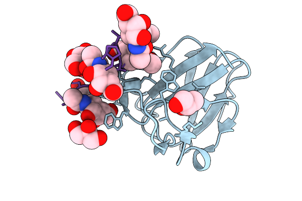
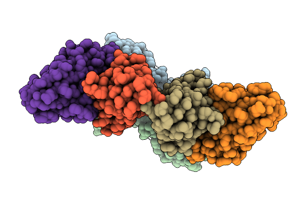
Organism: Escherichia coli, Synthetic construct
Method: X-RAY DIFFRACTION Release Date: 2025-12-24 Classification: SUGAR BINDING PROTEIN Ligands: A2G |
 |
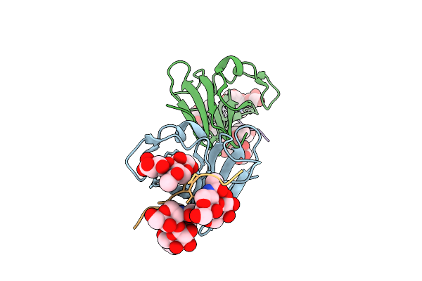
Organism: Escherichia coli, Synthetic construct
Method: X-RAY DIFFRACTION Release Date: 2025-12-24 Classification: SUGAR BINDING PROTEIN Ligands: GOL, A2G |
 |
Organism: Escherichia coli, Synthetic construct
Method: X-RAY DIFFRACTION Release Date: 2025-12-24 Classification: SUGAR BINDING PROTEIN |
 |
Organism: Homo sapiens
Method: ELECTRON MICROSCOPY Release Date: 2025-12-03 Classification: METAL BINDING PROTEIN Ligands: ZN |
 |
Organism: Homo sapiens
Method: ELECTRON MICROSCOPY Release Date: 2025-12-03 Classification: SIGNALING PROTEIN Ligands: 6LT, ZN |
 |
Organism: Homo sapiens
Method: ELECTRON MICROSCOPY Release Date: 2025-12-03 Classification: SIGNALING PROTEIN Ligands: 6LT, ZN |
 |
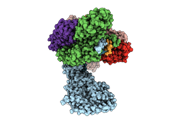
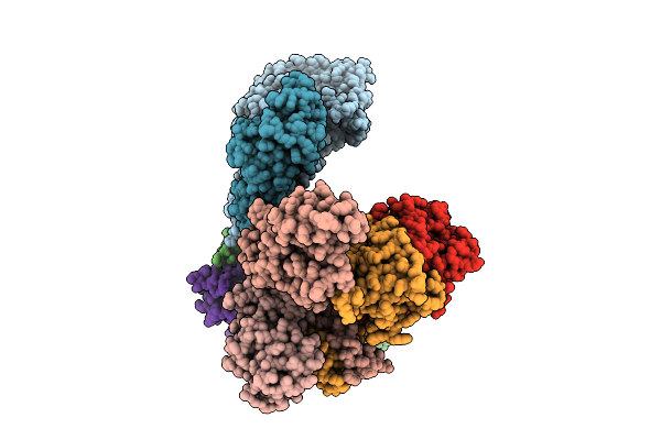
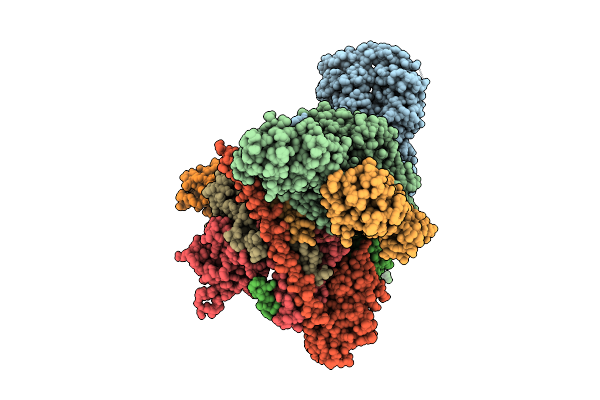
Cryo-Em Structure Of Cop9 Signalosome Precatalytic State With Neddylated Cullin-1
Organism: Homo sapiens
Method: ELECTRON MICROSCOPY Release Date: 2025-12-03 Classification: SIGNALING PROTEIN Ligands: ZN |
 |
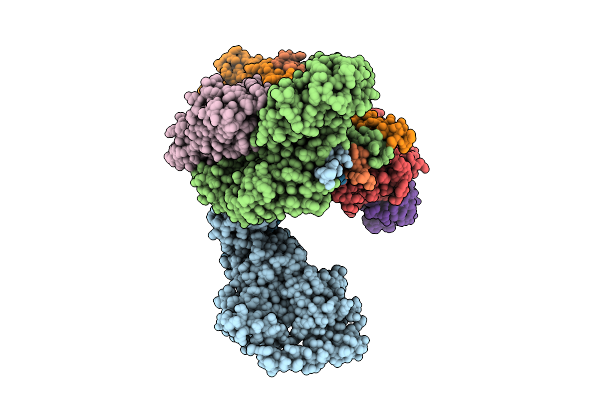
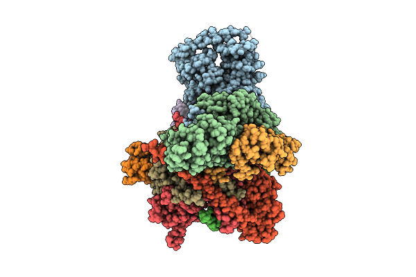
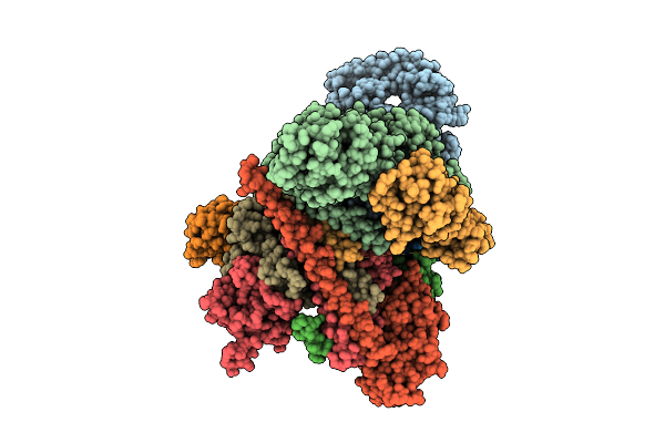
Cryo-Em Structure Of Cop9 Signalosome Precatalytic State With Neddylated Cullin-2
Organism: Homo sapiens
Method: ELECTRON MICROSCOPY Release Date: 2025-12-03 Classification: SIGNALING PROTEIN Ligands: ZN |
 |
Organism: Homo sapiens
Method: ELECTRON MICROSCOPY Release Date: 2025-12-03 Classification: SIGNALING PROTEIN Ligands: 6LT, ZN |
 |
Organism: Homo sapiens
Method: ELECTRON MICROSCOPY Release Date: 2025-12-03 Classification: SIGNALING PROTEIN Ligands: ZN |
 |
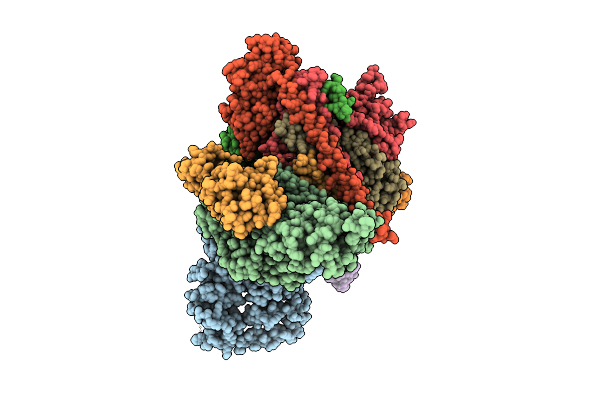
Cryo-Em Structure Of Cop9 Signalosome Precatalytic State With Neddylated Cullin-4A
Organism: Homo sapiens
Method: ELECTRON MICROSCOPY Release Date: 2025-12-03 Classification: SIGNALING PROTEIN Ligands: ZN |
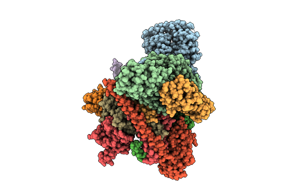 |
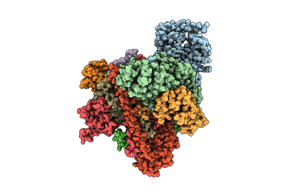
Cryo-Em Structure Of Cop9 Signalosome Precatalytic State With Neddylated Cullin-3
Organism: Homo sapiens
Method: ELECTRON MICROSCOPY Release Date: 2025-12-03 Classification: SIGNALING PROTEIN Ligands: ZN |
 |
Organism: Homo sapiens
Method: ELECTRON MICROSCOPY Release Date: 2025-12-03 Classification: SIGNALING PROTEIN Ligands: ZN, A1CH3 |
 |
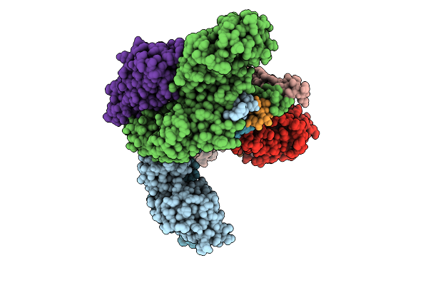
Cryo-Em Structure Of Human Complement C1S Cub Domain In Complex With Ray121
Organism: Homo sapiens
Method: ELECTRON MICROSCOPY Release Date: 2025-11-19 Classification: IMMUNE SYSTEM Ligands: CA |
 |
Organism: Homo sapiens
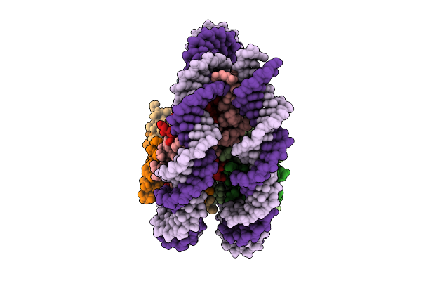
Method: ELECTRON MICROSCOPY Release Date: 2025-11-05 Classification: DNA BINDING PROTEIN |
 |
Organism: Homo sapiens
Method: ELECTRON MICROSCOPY Release Date: 2025-11-05 Classification: DNA BINDING PROTEIN |
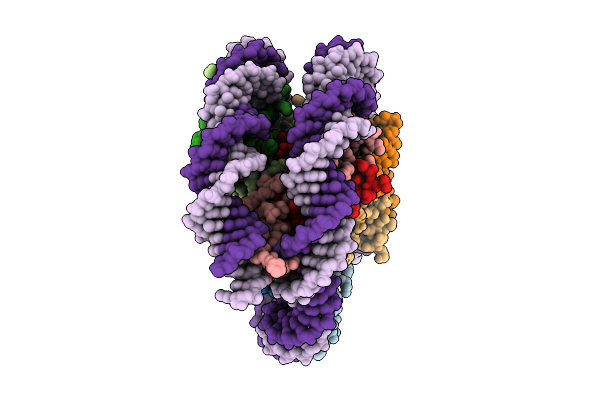
 |
Organism: Homo sapiens
Method: ELECTRON MICROSCOPY Release Date: 2025-11-05 Classification: DNA BINDING PROTEIN |
 |
Organism: Homo sapiens
Method: ELECTRON MICROSCOPY Release Date: 2025-11-05 Classification: DNA BINDING PROTEIN |
 |
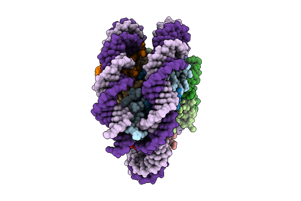
Organism: Homo sapiens, Synthetic construct
Method: ELECTRON MICROSCOPY Release Date: 2025-11-05 Classification: DNA BINDING PROTEIN |
 |
Organism: Homo sapiens, Synthetic construct
Method: ELECTRON MICROSCOPY Release Date: 2025-11-05 Classification: DNA BINDING PROTEIN |