
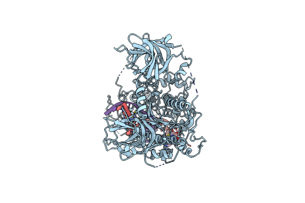
Search Count: 84
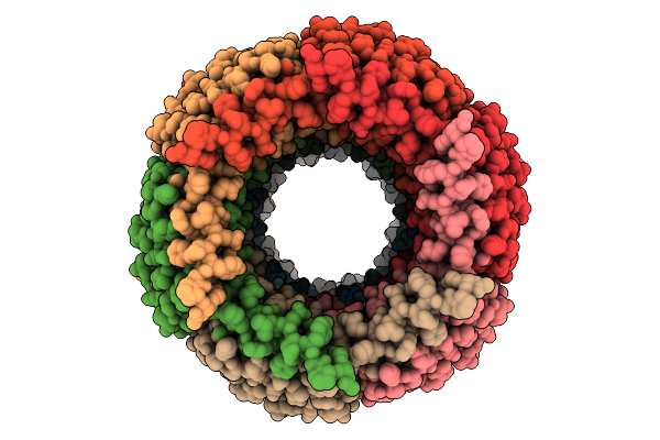 |
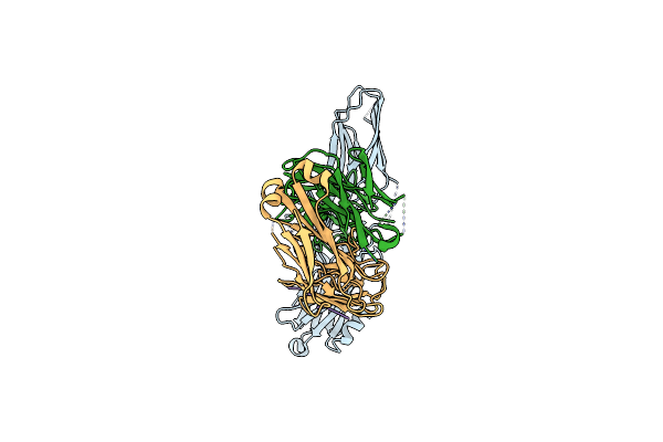
Organism: Escherichia phage p1
Method: ELECTRON MICROSCOPY Release Date: 2025-09-10 Classification: VIRAL PROTEIN |
 |
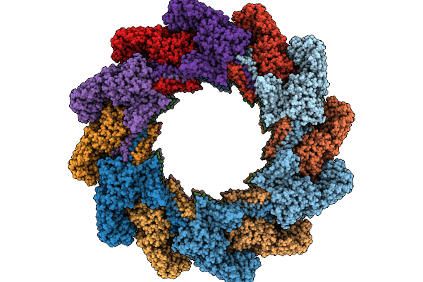
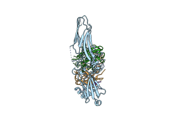
Organism: Escherichia phage p1
Method: ELECTRON MICROSCOPY Release Date: 2025-09-10 Classification: VIRAL PROTEIN |
 |
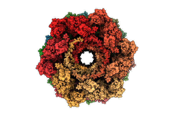
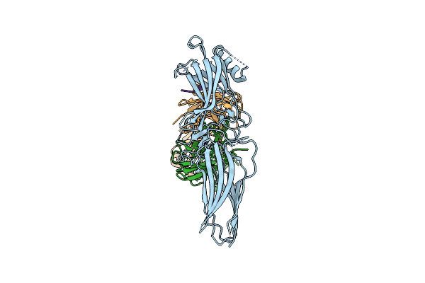
Organism: Escherichia phage p1
Method: ELECTRON MICROSCOPY Release Date: 2025-09-10 Classification: VIRAL PROTEIN |
 |
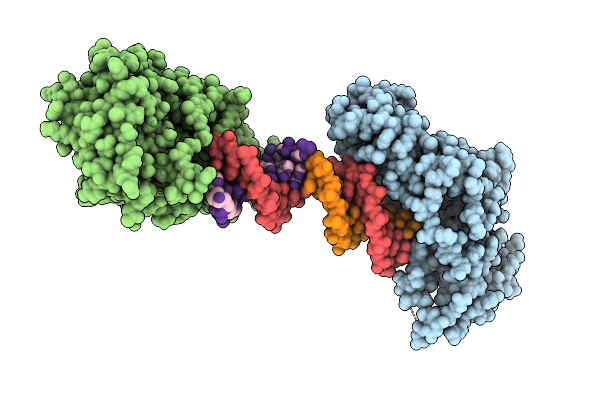
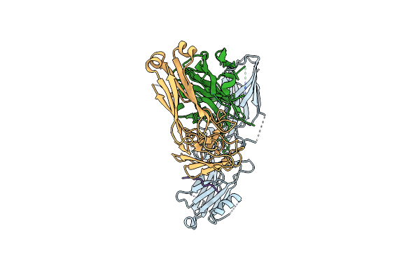
Organism: Escherichia phage p1
Method: ELECTRON MICROSCOPY Release Date: 2025-09-10 Classification: VIRAL PROTEIN |
 |
Organism: Zea mays, Synthetic construct
Method: X-RAY DIFFRACTION Release Date: 2025-07-16 Classification: PLANT PROTEIN/DNA Ligands: MG |
 |
Organism: Zea mays
Method: X-RAY DIFFRACTION Release Date: 2025-07-16 Classification: PLANT PROTEIN Ligands: MN |
 |
Organism: Bos taurus, Homo sapiens, Synthetic construct
Method: ELECTRON MICROSCOPY Release Date: 2025-04-02 Classification: SIGNALING PROTEIN/IMMUNE SYSTEM Ligands: CLR |
 |
Organism: Bos taurus
Method: ELECTRON MICROSCOPY Release Date: 2025-03-05 Classification: BIOSYNTHETIC PROTEIN |
 |
Organism: Bos taurus, Synthetic construct, Homo sapiens
Method: ELECTRON MICROSCOPY Release Date: 2025-01-22 Classification: SIGNALING PROTEIN/IMMUNE SYSTEM |
 |
Human Ackr3 Phosphorylated By Grk5 In Complex With Arrestin3 Reconstructed Without Receptor/Micelle
Organism: Synthetic construct, Homo sapiens, Bos taurus
Method: ELECTRON MICROSCOPY Release Date: 2025-01-22 Classification: SIGNALING PROTEIN/IMMUNE SYSTEM |
 |
Human Ackr3 Phosphorylated By Grk2 In Complex With Arrestin3 Reconstructed Without Receptor/Micelle
Organism: Synthetic construct, Bos taurus, Homo sapiens
Method: ELECTRON MICROSCOPY Release Date: 2025-01-22 Classification: SIGNALING PROTEIN/IMMUNE SYSTEM |
 |
Human Ackr3 With C Tail Extended By 12 Glycines Phosphorylated By Grk5 In Complex With Arrestin2 Reconstructed Without Receptor/Micelle
Organism: Bos taurus, Synthetic construct, Homo sapiens
Method: ELECTRON MICROSCOPY Release Date: 2025-01-22 Classification: SIGNALING PROTEIN/IMMUNE SYSTEM |
 |
Cryoem Structure Of Human Ackr3 Phosphorylated By Grk5 In Complex With Arrestin3 Variant With The C Edge Loop From Arrestin2 Inserted
Organism: Synthetic construct, Homo sapiens, Bos taurus
Method: ELECTRON MICROSCOPY Release Date: 2025-01-22 Classification: SIGNALING PROTEIN/IMMUNE SYSTEM |
 |
Cryo-Em Structure Of Human Dnmt1 (Aa:351-1616) In Complex With Ubiquitinated Paf15 And Hemimethylated Dna Analog
Organism: Homo sapiens, Synthetic construct
Method: ELECTRON MICROSCOPY Release Date: 2025-01-15 Classification: TRANSFERASE Ligands: SAH, ZN |
 |
E. Coli 70S Ribosome Complexed With P. Putida Trnaile2 At The A-Site And P-Site
Organism: Escherichia coli, Pseudomonas putida nbrc 14164
Method: ELECTRON MICROSCOPY Release Date: 2024-11-06 Classification: RIBOSOME Ligands: MG |
 |
Organism: Escherichia coli, Escherichia coli bw25113, Pseudomonas putida nbrc 14164
Method: ELECTRON MICROSCOPY Release Date: 2024-11-06 Classification: RIBOSOME Ligands: MG |
 |
Organism: Escherichia coli, Escherichia coli bw25113, Pseudomonas putida nbrc 14164
Method: ELECTRON MICROSCOPY Release Date: 2024-11-06 Classification: RIBOSOME Ligands: MG |
 |
Organism: Escherichia coli, Escherichia coli bw25113, Pseudomonas putida nbrc 14164
Method: ELECTRON MICROSCOPY Release Date: 2024-11-06 Classification: RIBOSOME Ligands: MG |
 |
Organism: Escherichia coli, Escherichia coli bw25113, Pseudomonas putida nbrc 14164
Method: ELECTRON MICROSCOPY Release Date: 2024-11-06 Classification: RIBOSOME Ligands: MG |
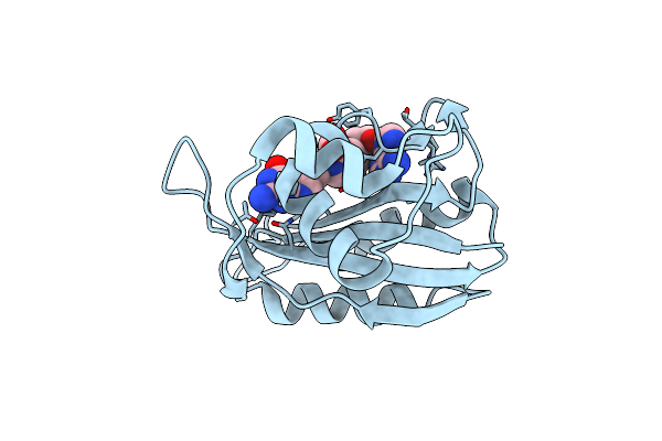 |
Crystal Structure Of Escherichia Coli Hppk In Complex With Bisubstrate Inhibitor Hp-101
Organism: Escherichia coli
Method: X-RAY DIFFRACTION Resolution:1.70 Å Release Date: 2024-07-03 Classification: TRANSFERASE/INHIBITOR Ligands: ZX4 |