Search Count: 269
 |
Molecular Basis Of Pathogenicity Of The Recently Emerged Fcov-23 Coronavirus. Complex Of Fapn With Fcov-23 Rbd
Organism: Felis catus, Feline coronavirus
Method: ELECTRON MICROSCOPY Release Date: 2025-07-09 Classification: VIRAL PROTEIN/HYDROLASE Ligands: NAG, ZN |
 |
Molecular Basis Of Pathogenicity Of The Recently Emerged Fcov-23 Coronavirus. Fcov-23 S Short
Organism: Feline coronavirus
Method: ELECTRON MICROSCOPY Release Date: 2025-07-09 Classification: VIRAL PROTEIN Ligands: NAG, PAM |
 |
Molecular Basis Of Pathogenicity Of The Recently Emerged Fcov-23 Coronavirus. Fcov-23 S Do In Proximal Conformation (Local Refinement)
Organism: Feline coronavirus
Method: ELECTRON MICROSCOPY Release Date: 2025-07-09 Classification: VIRAL PROTEIN Ligands: NAG |
 |
Molecular Basis Of Pathogenicity Of The Recently Emerged Fcov-23 Coronavirus. Fcov-23 S Long With Do In Swung-Out Conformation
Organism: Feline coronavirus
Method: ELECTRON MICROSCOPY Release Date: 2025-07-09 Classification: VIRAL PROTEIN Ligands: NAG, PAM |
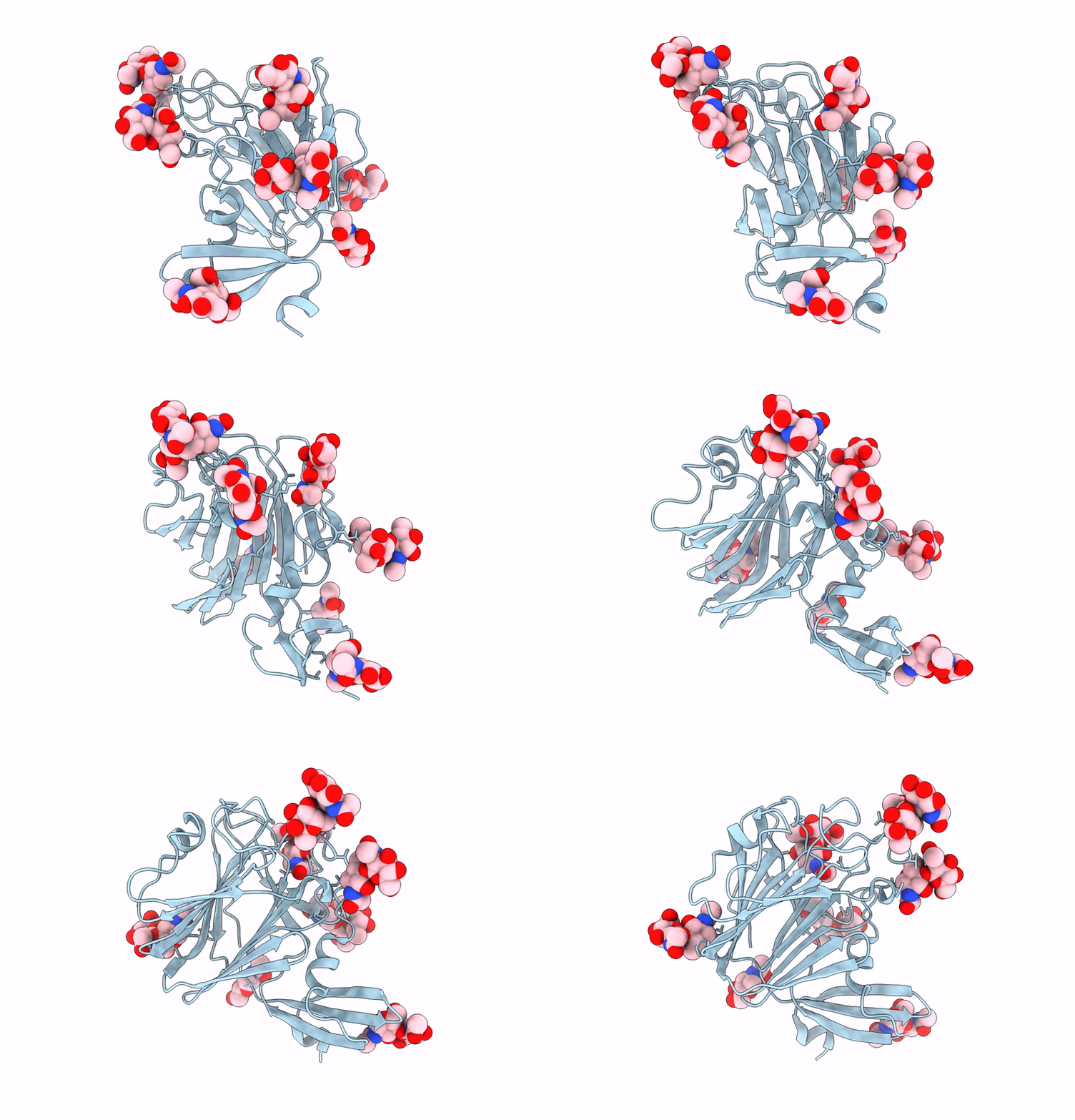 |
Molecular Basis Of Pathogenicity Of The Recently Emerged Fcov-23 Coronavirus. Fcov-23 S Long Domain 0 In Swung-Out Conformation (Local Refinement)
Organism: Feline coronavirus
Method: ELECTRON MICROSCOPY Release Date: 2025-07-09 Classification: VIRAL PROTEIN Ligands: NAG |
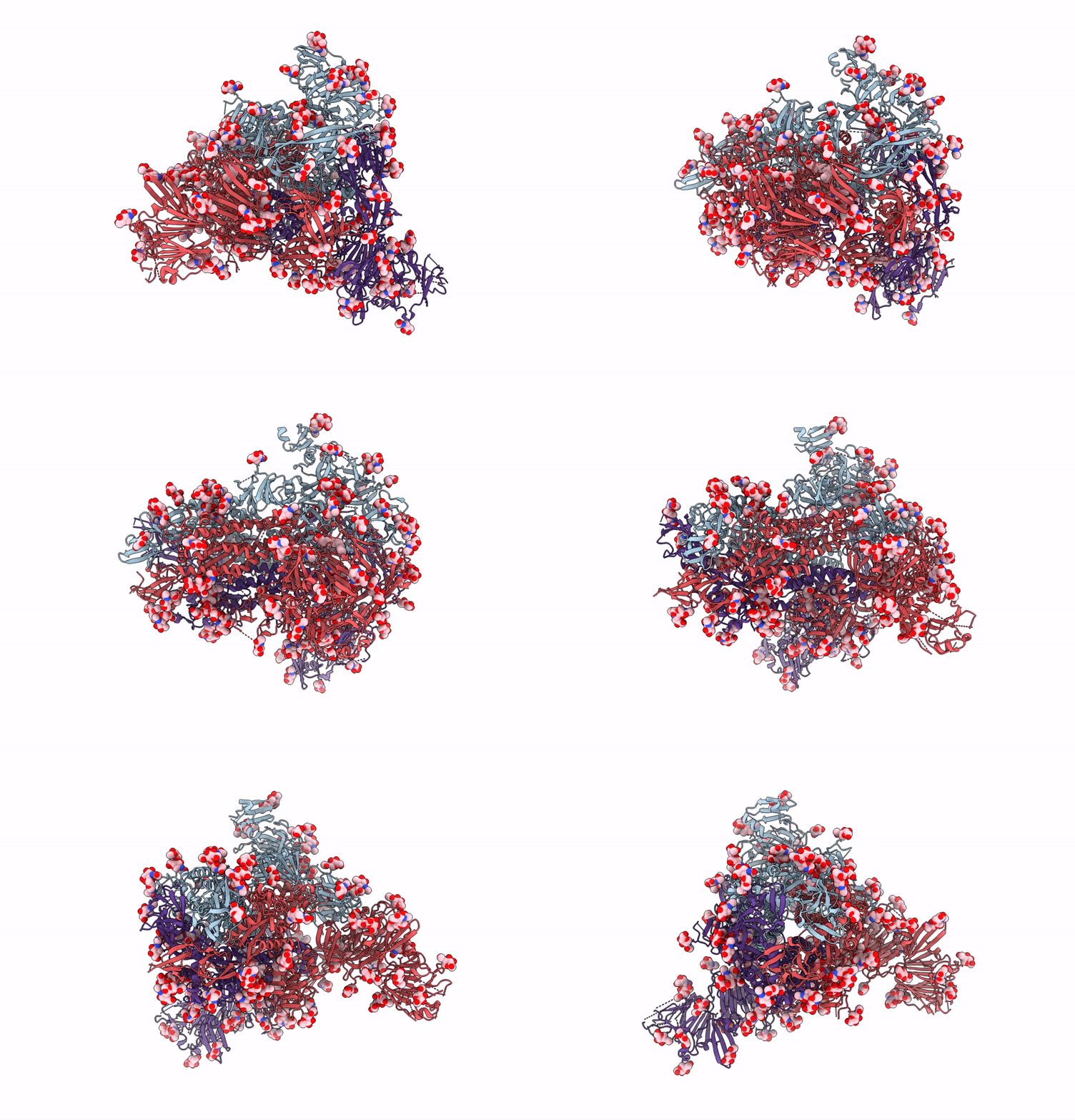 |
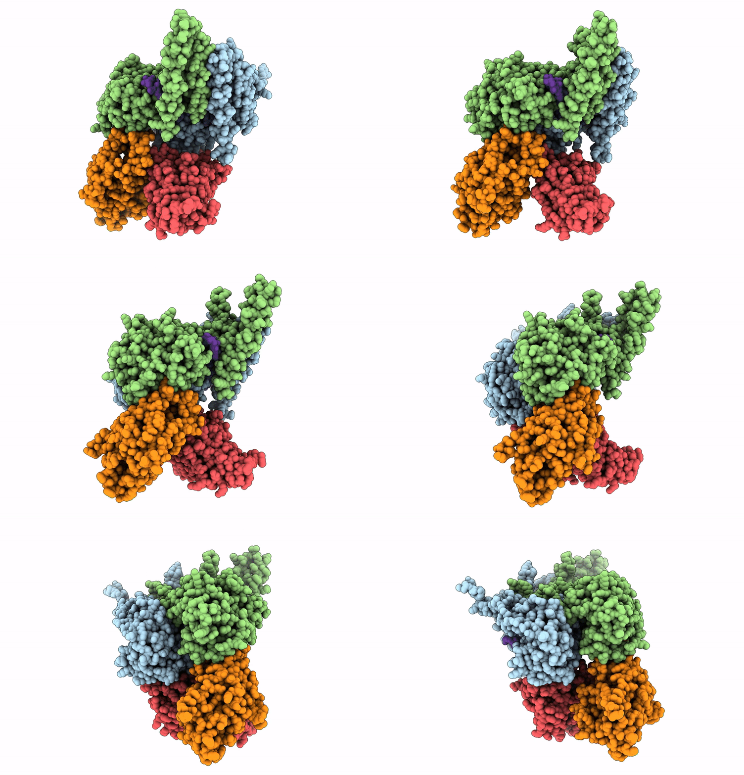
Molecular Basis Of Pathogenicity Of The Recently Emerged Fcov-23 Coronavirus. Fcov-23 S Long With Do In Mixed Conformations (Global Refinement).
Organism: Feline coronavirus
Method: ELECTRON MICROSCOPY Release Date: 2025-07-09 Classification: VIRAL PROTEIN Ligands: NAG, PAM |
 |
Organism: Zaire ebolavirus, Homo sapiens
Method: ELECTRON MICROSCOPY Release Date: 2025-01-29 Classification: VIRAL PROTEIN/RNA |
 |
Organism: Homo sapiens
Method: X-RAY DIFFRACTION Resolution:1.62 Å Release Date: 2024-12-04 Classification: HYDROLASE Ligands: MN, A1H0B |
 |
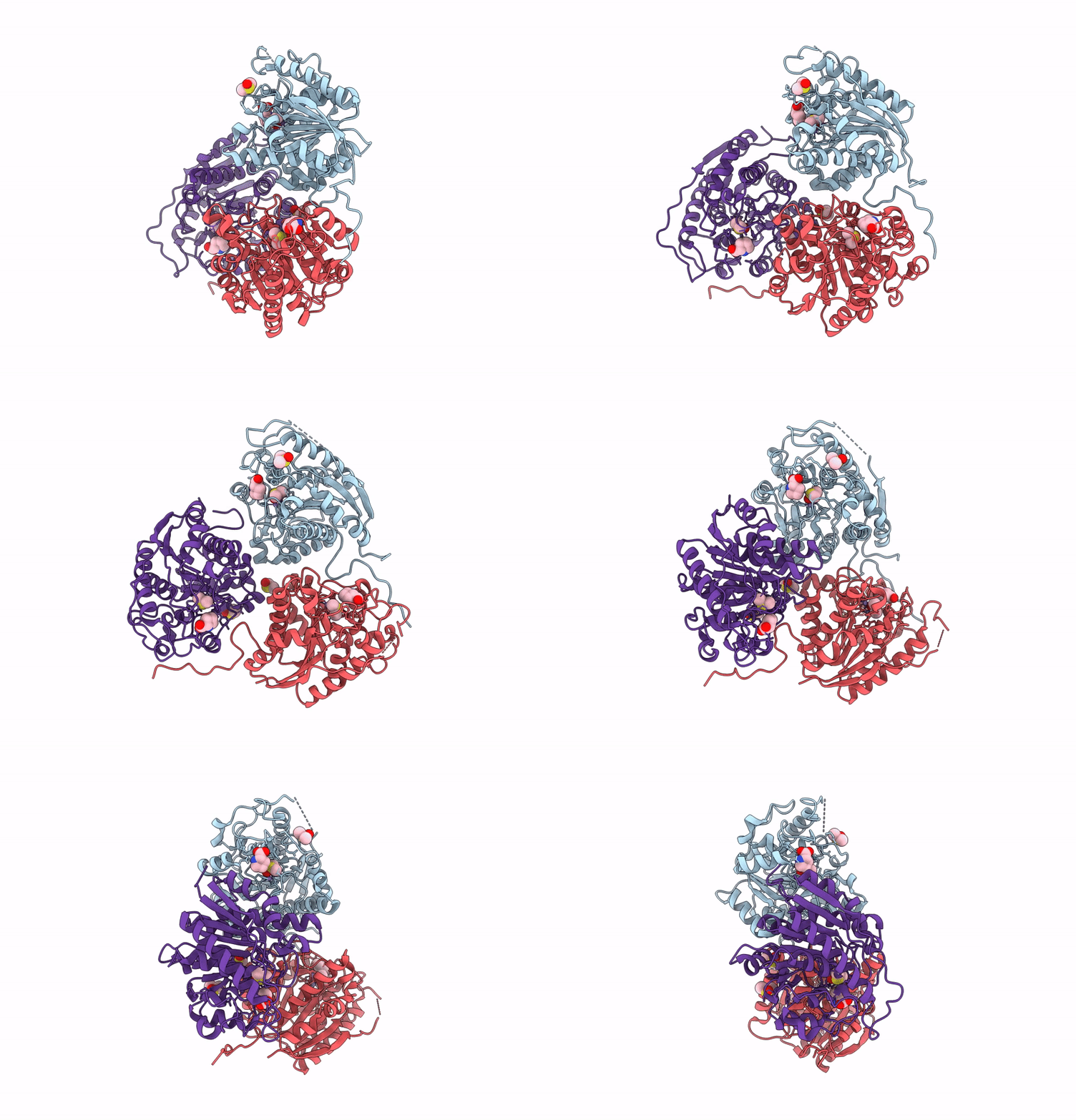
Organism: Homo sapiens
Method: X-RAY DIFFRACTION Resolution:1.86 Å Release Date: 2024-12-04 Classification: HYDROLASE Ligands: MN, A1H0A |
 |
Organism: Homo sapiens
Method: X-RAY DIFFRACTION Resolution:1.90 Å Release Date: 2024-12-04 Classification: HYDROLASE Ligands: MN, DMS, PRO |
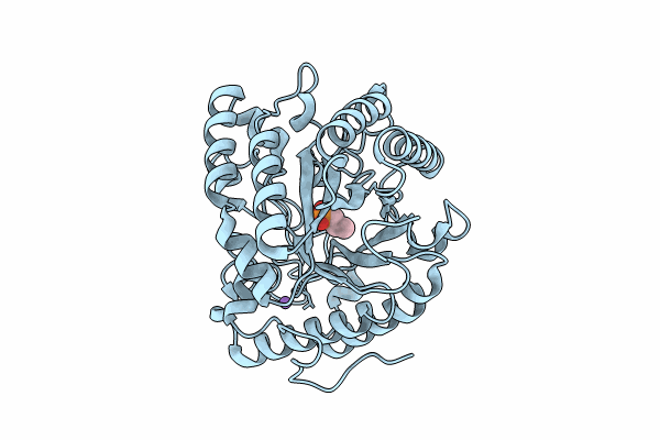 |
Crystal Structure Of Apo- Exo-Beta-(1,3)-Glucanase From Aspergillus Oryzae (Aobgl)
Organism: Aspergillus oryzae
Method: X-RAY DIFFRACTION Resolution:1.75 Å Release Date: 2024-11-06 Classification: HYDROLASE Ligands: A1L0T, NA |
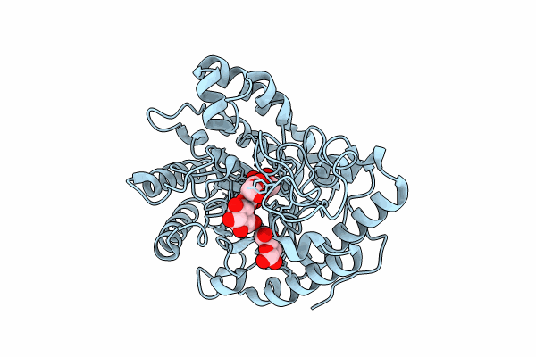 |
Crystal Structure Of Exo-Beta-(1,3)-Glucanase From Aspergillus Oryzae (Aobgl) As A Complex With Cellobiose
Organism: Aspergillus oryzae
Method: X-RAY DIFFRACTION Resolution:1.73 Å Release Date: 2024-11-06 Classification: HYDROLASE Ligands: BGC, NA |
 |
High-Resolution Crystal Structure Of Exo-Beta-(1,3)-Glucanase From Aspergillus Oryzae (Aobgl) As A Complex With Glucose
Organism: Aspergillus oryzae
Method: X-RAY DIFFRACTION Resolution:1.20 Å Release Date: 2024-11-06 Classification: HYDROLASE Ligands: EDO, GOL, BGC, CL, NA |
 |
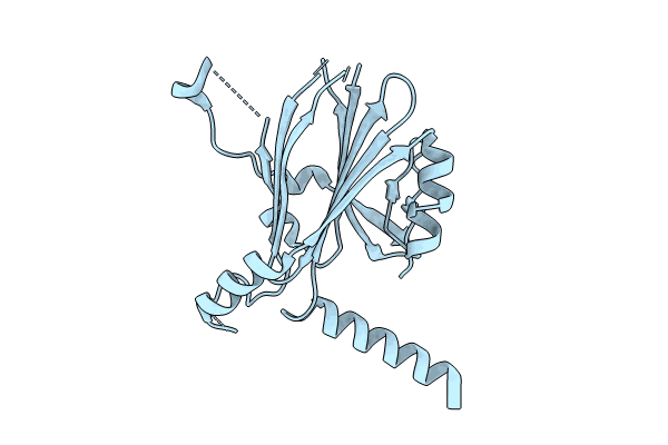
Organism: Bombyx mori cytoplasmic polyhedrosis virus, Homo sapiens
Method: X-RAY DIFFRACTION Resolution:2.55 Å Release Date: 2024-06-05 Classification: VIRAL PROTEIN |
 |
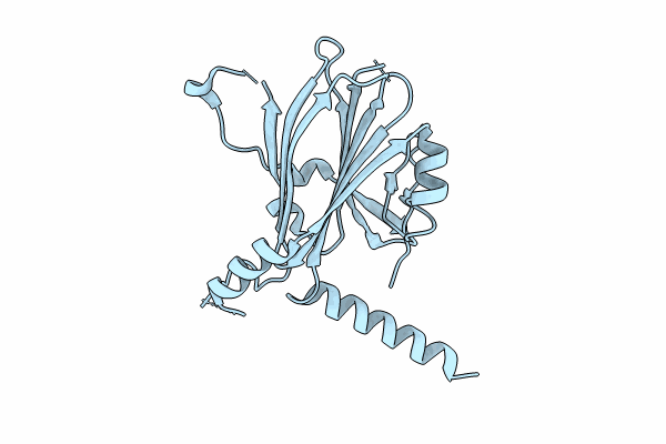
Organism: Bombyx mori cypovirus 1, Homo sapiens
Method: X-RAY DIFFRACTION Resolution:2.04 Å Release Date: 2024-06-05 Classification: VIRAL PROTEIN |
 |
Organism: Bombyx mori cypovirus 1, Homo sapiens
Method: X-RAY DIFFRACTION Resolution:2.00 Å Release Date: 2024-06-05 Classification: VIRAL PROTEIN |
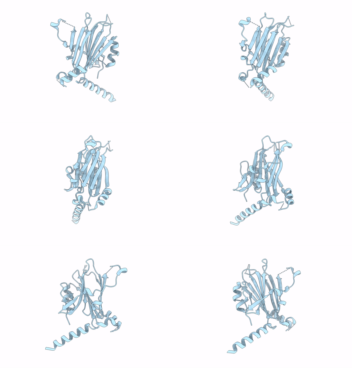 |
Organism: Bombyx mori cytoplasmic polyhedrosis virus, Homo sapiens
Method: X-RAY DIFFRACTION Resolution:1.92 Å Release Date: 2024-04-17 Classification: VIRAL PROTEIN |
 |
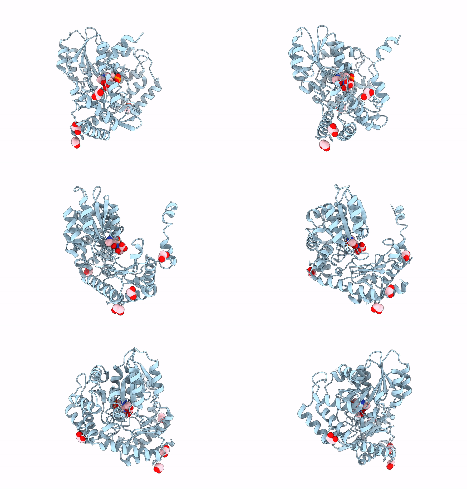
Crystal Structure Of Serine Palmitoyltransferase Soaked In 190 Mm D-Serine Solution
Organism: Sphingobacterium multivorum
Method: X-RAY DIFFRACTION Resolution:1.65 Å Release Date: 2024-04-10 Classification: TRANSFERASE Ligands: PLS, EDO |
 |
Crystal Structure Of Serine Palmitoyltransferase Complexed With D-Methylserine
Organism: Sphingobacterium multivorum
Method: X-RAY DIFFRACTION Resolution:1.70 Å Release Date: 2024-04-10 Classification: TRANSFERASE Ligands: S5R, EDO |
 |
Crystal Structure Of The Collagen Binding Domain Of Cnm From Streptococcus Mutans
Organism: Streptococcus mutans
Method: X-RAY DIFFRACTION Resolution:1.81 Å Release Date: 2024-04-10 Classification: PROTEIN BINDING Ligands: SO4, GOL |

