Search Count: 162
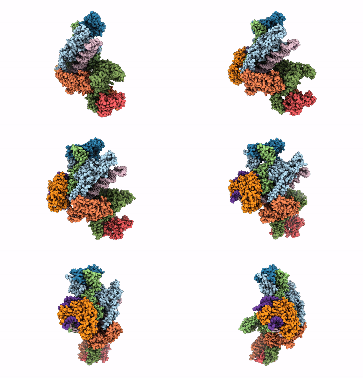 |
Organism: Escherichia coli
Method: ELECTRON MICROSCOPY Release Date: 2025-07-02 Classification: ANTIVIRAL PROTEIN Ligands: ADP, ZN |
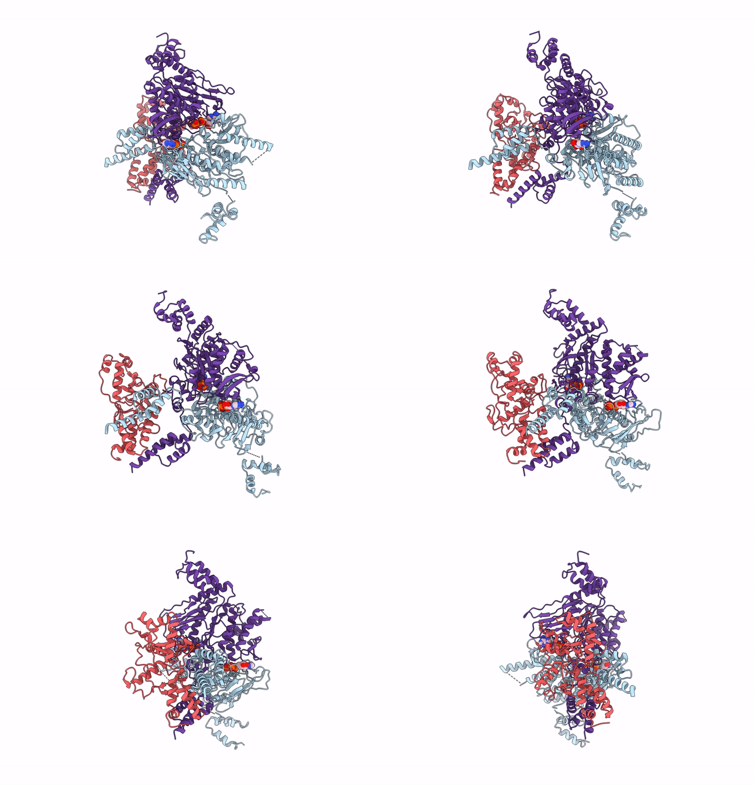 |
Organism: Escherichia coli
Method: ELECTRON MICROSCOPY Release Date: 2025-07-02 Classification: ANTIVIRAL PROTEIN Ligands: ZN, ATP |
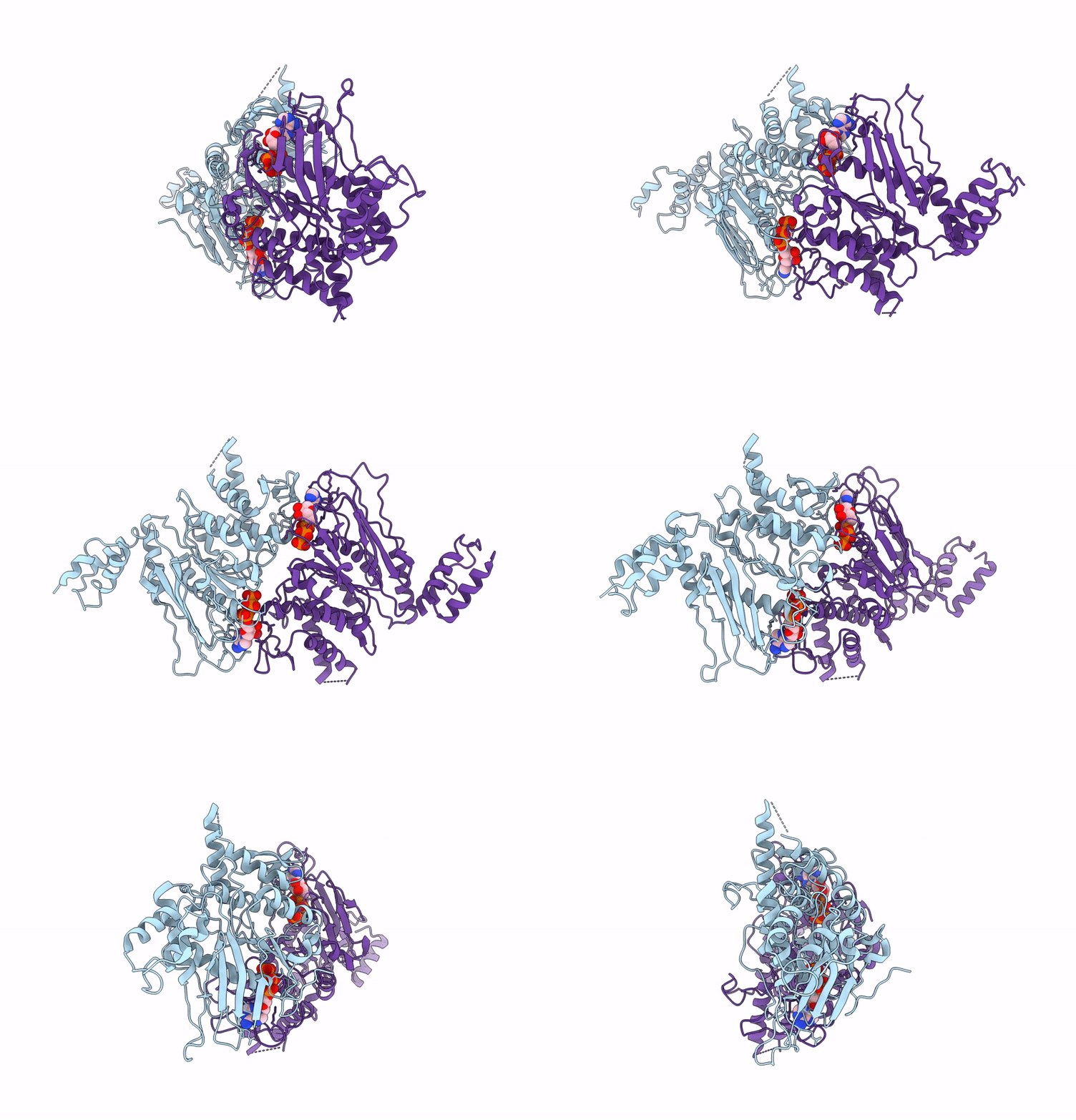 |
Organism: Escherichia coli
Method: ELECTRON MICROSCOPY Release Date: 2025-07-02 Classification: ANTIVIRAL PROTEIN Ligands: ATP |
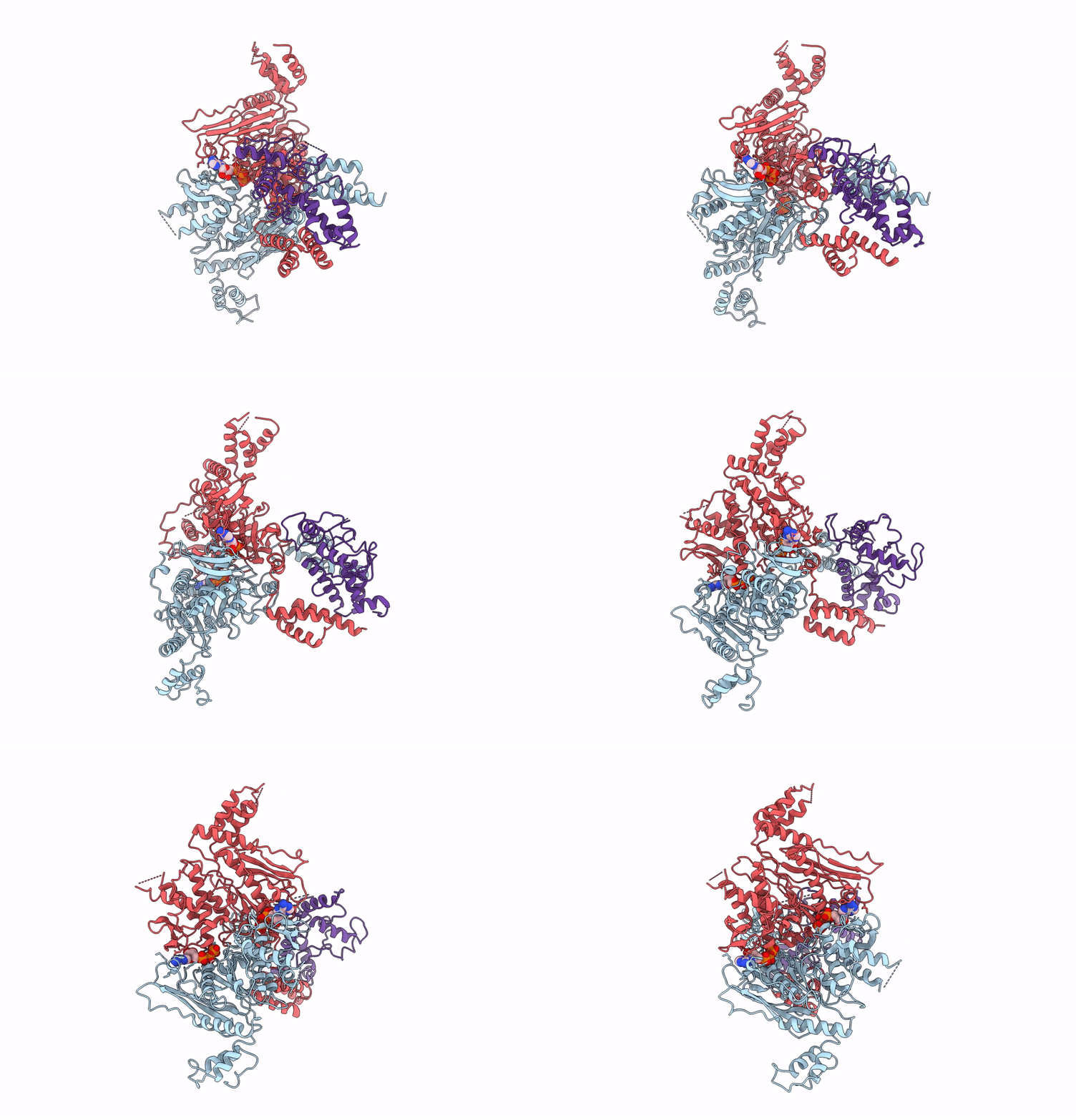 |
Organism: Escherichia coli
Method: ELECTRON MICROSCOPY Release Date: 2025-07-02 Classification: IMMUNE SYSTEM Ligands: ATP, ZN |
 |
Organism: Bacillus sp. hmf5848
Method: ELECTRON MICROSCOPY Release Date: 2025-06-04 Classification: ANTIVIRAL PROTEIN |
 |
Organism: Bacillus sp. hmf5848
Method: ELECTRON MICROSCOPY Release Date: 2025-06-04 Classification: ANTIVIRAL PROTEIN |
 |
Organism: Bacillus sp. hmf5848
Method: ELECTRON MICROSCOPY Release Date: 2025-06-04 Classification: ANTIVIRAL PROTEIN |
 |
Organism: Bacillus sp. hmf5848
Method: ELECTRON MICROSCOPY Release Date: 2025-06-04 Classification: ANTIVIRAL PROTEIN |
 |
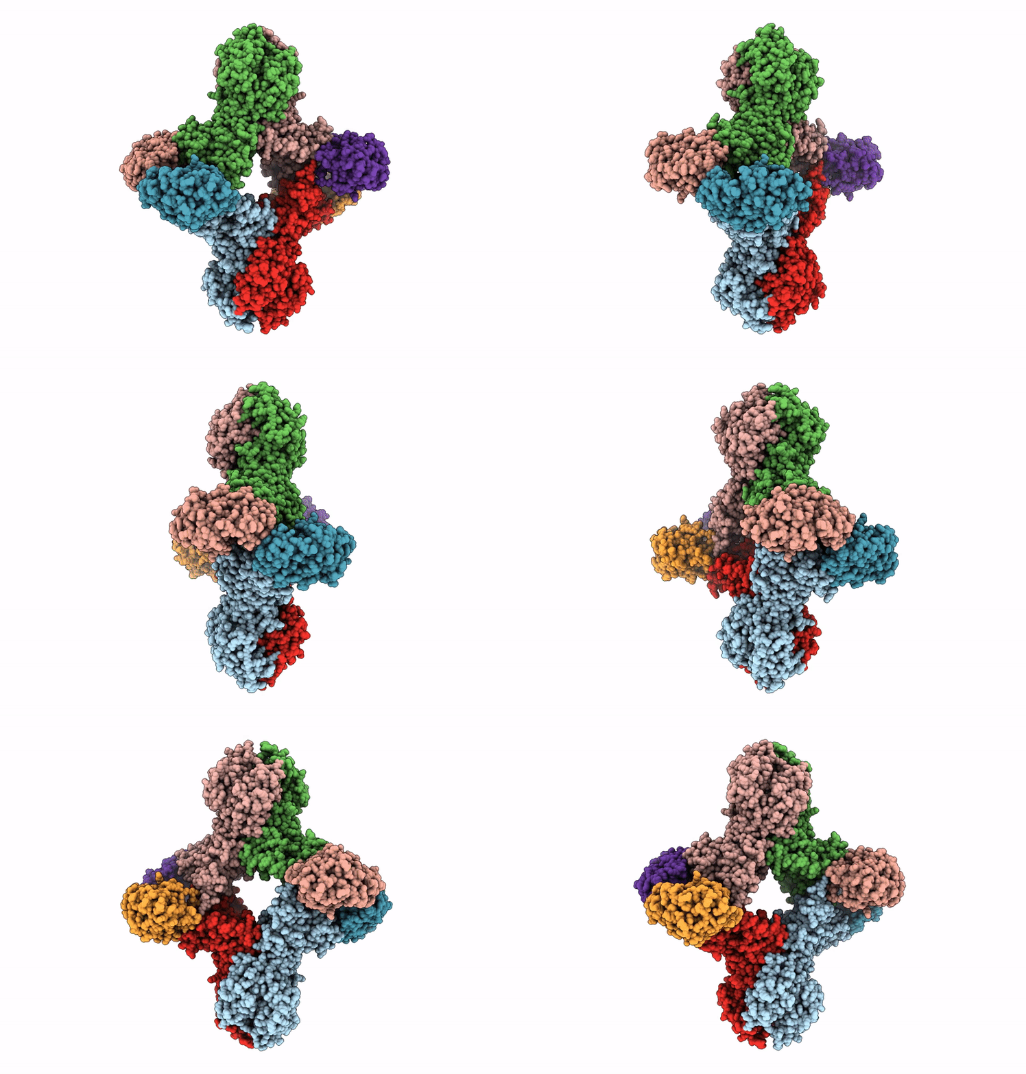
Molecular Basis For Hera-Duf Supramolecular Complex In Anti-Phage Defense - Assembly 3
Organism: Bacillus sp. hmf5848
Method: ELECTRON MICROSCOPY Release Date: 2025-06-04 Classification: ANTIVIRAL PROTEIN |
 |
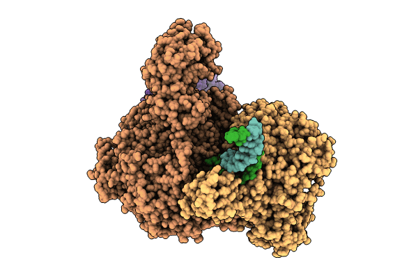
The Crystal Structure Of Styrene Monooxygenase Stya From Streptomyces Vilmorinianum
Organism: Streptomyces vilmorinianum
Method: X-RAY DIFFRACTION Resolution:2.86 Å Release Date: 2025-03-19 Classification: OXIDOREDUCTASE |
 |
Organism: Vibrio cholerae
Method: ELECTRON MICROSCOPY Release Date: 2024-10-23 Classification: IMMUNE SYSTEM/DNA Ligands: MG |
 |
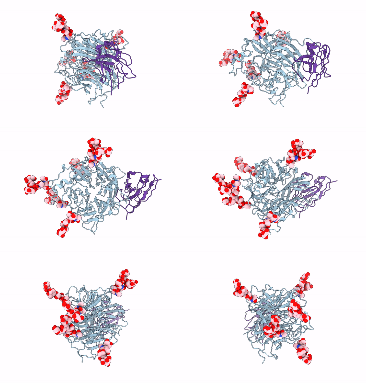
Structure Of Nipah Virus Bangladesh String G Protein Ectodomain Monomer Bound To Single-Domain Antibody N425 At 3.22 Angstroms Overall Resolution
Organism: Homo sapiens, Henipavirus nipahense
Method: ELECTRON MICROSCOPY Release Date: 2024-09-18 Classification: VIRAL PROTEIN |
 |
Structure Of Nipah Virus Malaysia String G Protein Ectodomain Monomer Bound To Single-Domain Antibody N425 At 3.63 Angstroms Overall Resolution
Organism: Henipavirus nipahense, Homo sapiens
Method: ELECTRON MICROSCOPY Release Date: 2024-09-18 Classification: VIRAL PROTEIN |
 |
Structure Of Nipah Virus Bangladesh String G Protein Ectodomain Tetramer Bound To Single-Domain Antibody N425 At 5.87 Angstroms Overall Resolution
Organism: Henipavirus nipahense, Homo sapiens
Method: ELECTRON MICROSCOPY Release Date: 2024-09-18 Classification: VIRAL PROTEIN |
 |
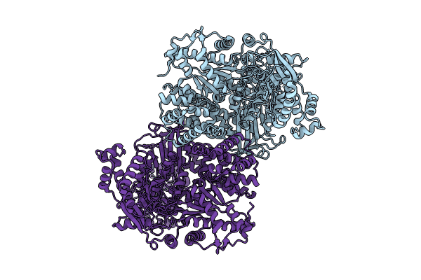
Organism: Vibrio cholerae
Method: ELECTRON MICROSCOPY Release Date: 2024-09-04 Classification: IMMUNE SYSTEM/DNA |
 |
Organism: Vibrio cholerae
Method: ELECTRON MICROSCOPY Release Date: 2024-08-21 Classification: IMMUNE SYSTEM |
 |
Organism: Vibrio cholerae
Method: ELECTRON MICROSCOPY Release Date: 2024-08-21 Classification: IMMUNE SYSTEM/DNA |
 |
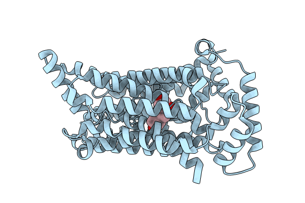
Cryo-Em Structure Of Human Urate Transporter Glut9 Bound To Substrate Urate
Organism: Homo sapiens
Method: ELECTRON MICROSCOPY Release Date: 2024-06-19 Classification: TRANSPORT PROTEIN Ligands: URC |
 |
Cryo-Em Structure Of Human Urate Transporter Glut9 Bound To Inhibitor Apigenin
Organism: Homo sapiens
Method: ELECTRON MICROSCOPY Release Date: 2024-06-19 Classification: TRANSPORT PROTEIN Ligands: AGI |
 |
Organism: Bacillus cereus
Method: ELECTRON MICROSCOPY Release Date: 2024-04-24 Classification: IMMUNE SYSTEM |

