
Search Count: 19
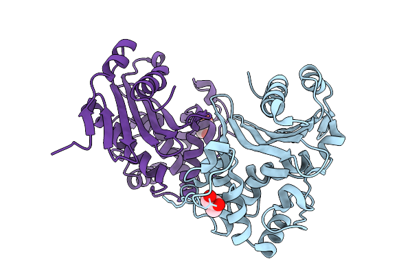 |
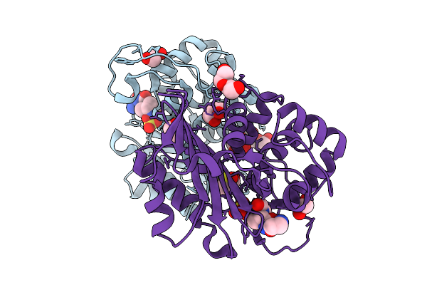
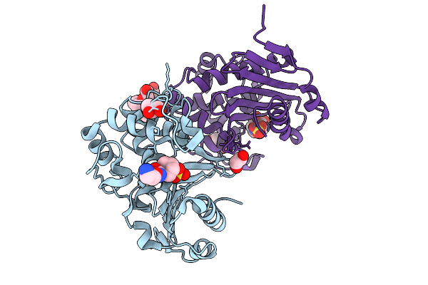
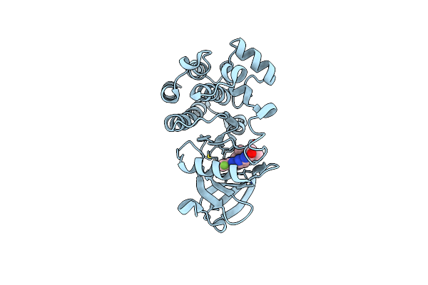
Organism: Klebsiella pneumoniae
Method: X-RAY DIFFRACTION Release Date: 2025-09-03 Classification: ANTIMICROBIAL PROTEIN Ligands: CL, GOL |
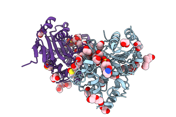 |
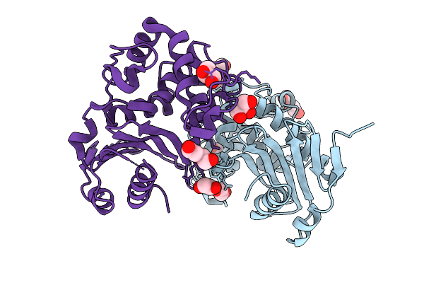
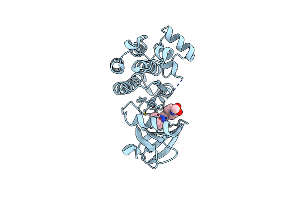
Organism: Klebsiella pneumoniae
Method: X-RAY DIFFRACTION Release Date: 2025-09-03 Classification: ANTIMICROBIAL PROTEIN Ligands: CL, PGE, PEG, EDO, OP0, DMS, GOL, PG4 |
 |
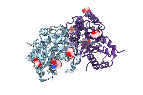
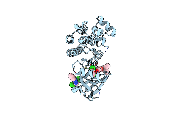
Organism: Enterobacter cloacae
Method: X-RAY DIFFRACTION Release Date: 2025-09-03 Classification: ANTIMICROBIAL PROTEIN Ligands: CL, GOL, EDO, PEG |
 |
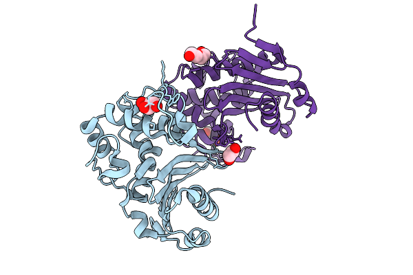
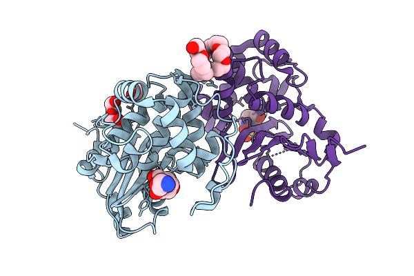
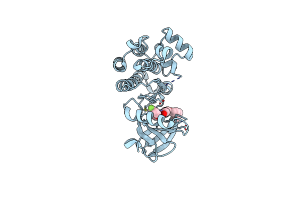
Organism: Enterobacter cloacae
Method: X-RAY DIFFRACTION Release Date: 2025-09-03 Classification: ANTIMICROBIAL PROTEIN Ligands: NXL, A1IY4, CL, EDO, 2PE |
 |
Organism: Enterobacter cloacae
Method: X-RAY DIFFRACTION Release Date: 2025-09-03 Classification: ANTIMICROBIAL PROTEIN Ligands: OP0, EDO, GOL, PEG, CL, DMS |
 |
Organism: Serratia marcescens
Method: X-RAY DIFFRACTION Release Date: 2025-09-03 Classification: ANTIMICROBIAL PROTEIN Ligands: PEG, CL, GOL, PG4, EDO |
 |
Organism: Serratia marcescens
Method: X-RAY DIFFRACTION Release Date: 2025-09-03 Classification: ANTIMICROBIAL PROTEIN Ligands: GOL, EDO, NXL, A1IY4, CL, PGE, PEG |
 |
Organism: Serratia marcescens
Method: X-RAY DIFFRACTION Release Date: 2025-09-03 Classification: ANTIMICROBIAL PROTEIN Ligands: OP0, PEG, EDO, A1IYS, CL, 2PE |
 |
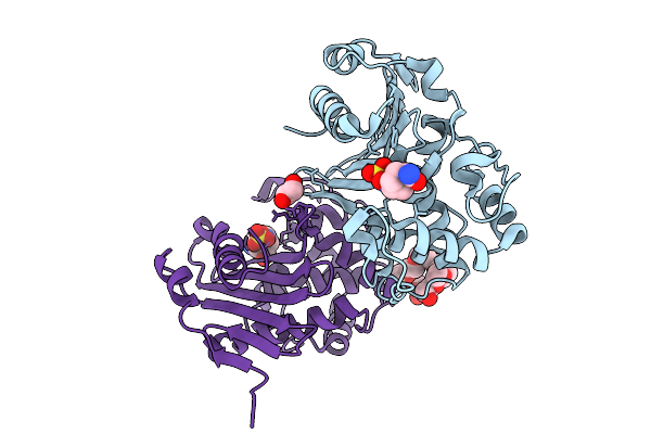
Organism: Enterobacter cloacae
Method: X-RAY DIFFRACTION Release Date: 2025-09-03 Classification: ANTIMICROBIAL PROTEIN Ligands: OP0, EDO, GOL, A1IYS, CL, 2PE |
 |
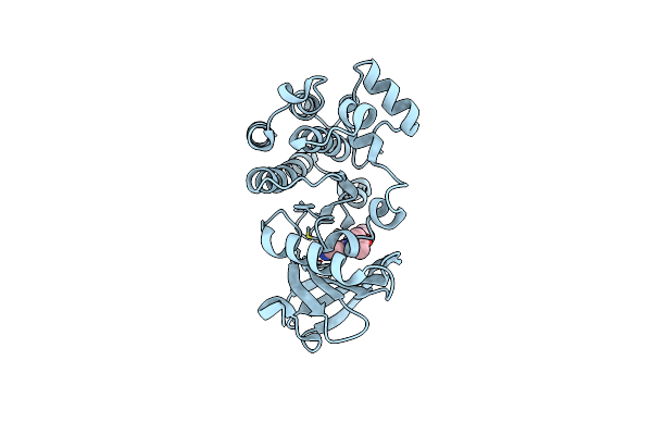
Crystal Structure Of A Radical Sam Methyltransferase From Sphaerobacter Thermophilus
Organism: Sphaerobacter thermophilus (strain dsm 20745 / s 6022)
Method: X-RAY DIFFRACTION Resolution:1.42 Å Release Date: 2019-04-10 Classification: TRANSFERASE Ligands: SF4, SAH, GOL, BR |
 |
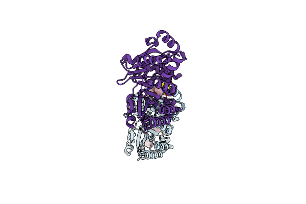
Structural Basis Of Mitochondrial Receptor Binding And Constriction By Dynamin-Related Protein 1
Organism: Homo sapiens
Method: ELECTRON MICROSCOPY Release Date: 2018-06-20 Classification: PROTEIN FIBRIL Ligands: GCP, MG |
 |
Bruton'S Tyrosine Kinase In Complex With A T-Butyl Cyanoacrylamide Inhibitor
Organism: Homo sapiens
Method: X-RAY DIFFRACTION Resolution:2.20 Å Release Date: 2015-05-13 Classification: TRANSFERASE Ligands: 4C9, NA, EDO, SO4, CL |
 |
Crystal Structure Of Btk Kinase Domain Complexed With 2-Isopropyl-7-(4-Methyl-Piperazin-1-Yl)-4-(5-Methyl-2H-Pyrazol-3-Ylamino)-2H-Phthalazin-1-One
Organism: Homo sapiens
Method: X-RAY DIFFRACTION Resolution:1.85 Å Release Date: 2011-01-12 Classification: TRANSFERASE/INHIBITOR Ligands: 027 |
 |
Organism: Homo sapiens
Method: X-RAY DIFFRACTION Resolution:2.55 Å Release Date: 2011-01-12 Classification: TRANSFERASE/INHIBITOR Ligands: 585 |
 |
Crystal Structure Of Btk Kinase Domain Complexed With (5-Amino-1-O-Tolyl-1H-Pyrazol-4-Yl)-[3-(1-Methanesulfonyl-Piperidin-4-Yl)-Phenyl]-Methanone
Organism: Homo sapiens
Method: X-RAY DIFFRACTION Resolution:2.21 Å Release Date: 2011-01-12 Classification: TRANSFERASE/INHIBITOR Ligands: 03C |
 |
Crystal Structure Of Btk Kinase Domain Complexed With 3-(2,6-Dichloro-Phenyl)-7-[4-(2-Diethylamino-Ethoxy)-Phenylamino]-1-Methyl-3,4-Dihydro-1H-Pyrimido[4,5-D]Pyrimidin-2-One
Organism: Homo sapiens
Method: X-RAY DIFFRACTION Resolution:2.00 Å Release Date: 2011-01-12 Classification: TRANSFERASE/INHIBITOR Ligands: LHL |
 |
Crystal Structure Of Btk Kinase Domain Complexed With 2-[4-(2-Diethylamino-Ethoxy)-Phenylamino]-6-(4-Fluoro-Phenoxy)-8-Methyl-8H-Pyrido[2,3-D]Pyrimidin-7-One
Organism: Homo sapiens
Method: X-RAY DIFFRACTION Resolution:1.75 Å Release Date: 2011-01-12 Classification: TRANSFERASE/INHIBITOR Ligands: 04K |
 |
Crystal Structure Of Btk Kinase Domain Complexed With 2-Methyl-5-[(E)-(3-Phenyl-Acryloyl)Amino]-N-(2-Phenyl-3H-Imidazo[4,5-B]Pyridin-6-Yl)-Benzamide
Organism: Homo sapiens
Method: X-RAY DIFFRACTION Resolution:1.85 Å Release Date: 2011-01-12 Classification: TRANSFERASE/INHIBITOR Ligands: 04L |
 |
Organism: Saccharomyces cerevisiae
Method: X-RAY DIFFRACTION Resolution:2.70 Å Release Date: 2010-10-27 Classification: PROTEIN BINDING |