
Search Count: 111
 |
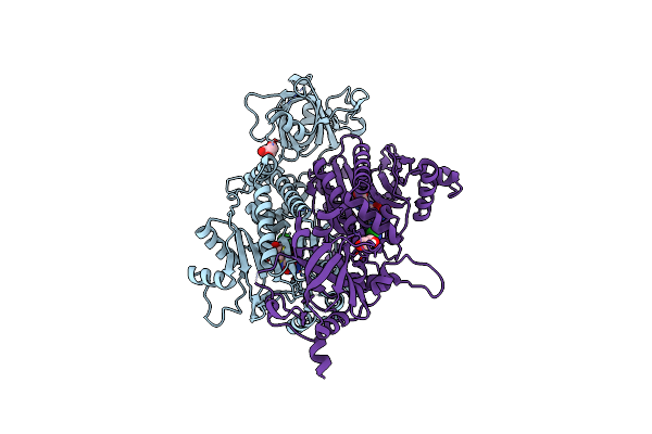
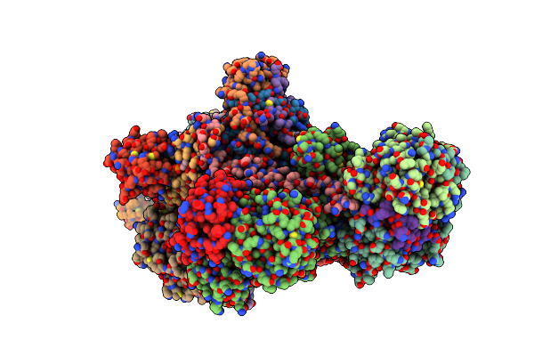
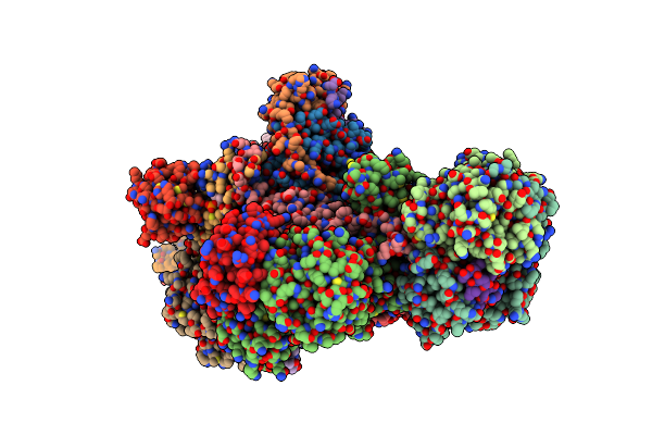
Crystal Structure Of Lysyl-Trna Synthetase From Plasmodium Falciparum Complexed With Lys-Ams
Organism: Plasmodium falciparum
Method: X-RAY DIFFRACTION Release Date: 2025-09-10 Classification: LIGASE Ligands: KAA |
 |
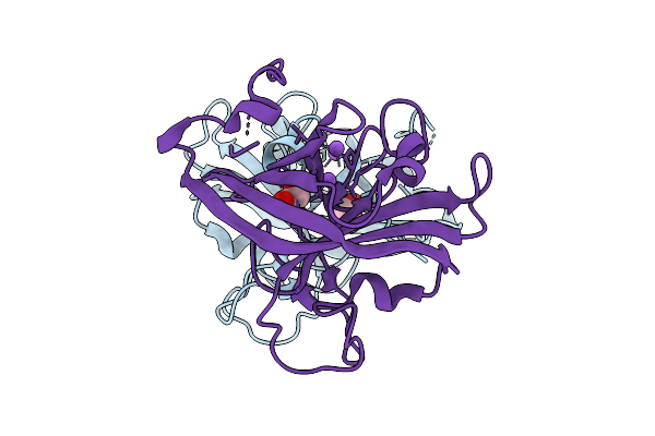
Organism: Thermobifida fusca yx
Method: X-RAY DIFFRACTION Release Date: 2025-08-06 Classification: HYDROLASE |
 |
Organism: Candidatus odinarchaeota
Method: ELECTRON MICROSCOPY Release Date: 2025-07-30 Classification: CELL CYCLE Ligands: G2P |
 |
Organism: Thermobifida fusca yx
Method: X-RAY DIFFRACTION Release Date: 2025-07-23 Classification: HYDROLASE Ligands: CL, NA, GOL |
 |
Crystal Structure Of C-Terminal Rigid Fragment Containing Middle And C-Terminal Domains Of The Basal Pilin Ebpb From Enterococcus Faecalis.
Organism: Enterococcus faecalis og1rf
Method: X-RAY DIFFRACTION Release Date: 2025-07-16 Classification: CELL ADHESION |
 |
Crystal Structure Of N-Terminal Flexible Domain Of The Shaft Pilin Ebpc From Enterococcus Faecalis.
Organism: Enterococcus faecalis og1rf
Method: X-RAY DIFFRACTION Release Date: 2025-07-16 Classification: CELL ADHESION |
 |
Crystal Structure Of N-Terminal Flexible Domain Of The Shaft Pilin Ebpc From Enterococcus Faecalis.
Organism: Enterococcus faecalis og1rf
Method: X-RAY DIFFRACTION Release Date: 2025-07-16 Classification: CELL ADHESION |
 |
Organism: Enterococcus faecalis og1rf
Method: X-RAY DIFFRACTION Release Date: 2025-07-16 Classification: CELL ADHESION |
 |
Crystal Structure Of C-Terminal Stable Fragment Of The Shaft Pilin Ebpc From Enterococcus Faecalis
Organism: Enterococcus faecalis og1rf
Method: X-RAY DIFFRACTION Release Date: 2025-07-16 Classification: CELL ADHESION Ligands: MG, NA |
 |
Crystal Structure Of The Basal Pilin Ebpb From Enterococcus Faecalis With A Partially Disordered N-Terminal Domain.
Organism: Enterococcus faecalis og1rf
Method: X-RAY DIFFRACTION Release Date: 2025-07-16 Classification: CELL ADHESION |
 |
Organism: Thermobifida fusca yx
Method: X-RAY DIFFRACTION Release Date: 2025-07-09 Classification: HYDROLASE Ligands: EDO, CL, NA |
 |
Organism: Plasmodium vivax
Method: X-RAY DIFFRACTION Release Date: 2025-06-04 Classification: LIGASE Ligands: SO4, EDO |
 |
Plasmodium Vivax Aspartyl-Trna Synthetase In Complex With Aspartyl-Adenylate (Asp-Amp) Complex.
Organism: Plasmodium vivax
Method: X-RAY DIFFRACTION Release Date: 2025-06-04 Classification: LIGASE Ligands: AMO, GOL, ACT |
 |
Plasmodium Vivax Aspartyl-Trna Synthetase In Complex With Aspartyl Sulfamoyl Adenosine (Asp-Ams) Complex
Organism: Plasmodium vivax
Method: X-RAY DIFFRACTION Release Date: 2025-06-04 Classification: LIGASE Ligands: DSZ, MG |
 |
Structural Studies Of Reaction Hijacking Inhibition Of A Malaria Parasite Aspartyl-Trna Synthetase.
Organism: Plasmodium vivax
Method: X-RAY DIFFRACTION Release Date: 2025-06-04 Classification: LIGASE Ligands: A1B0L, GOL |
 |
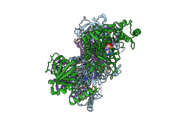
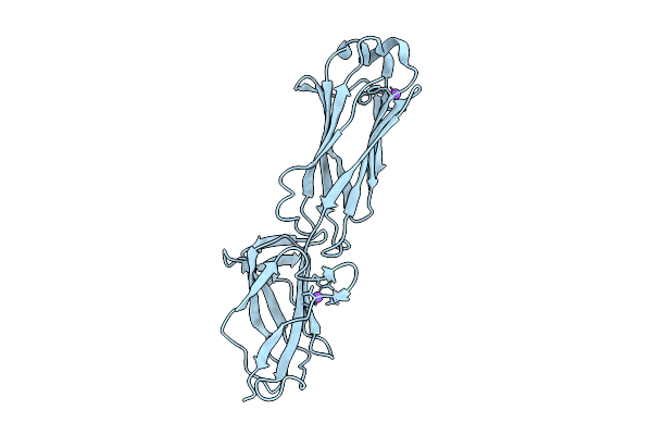
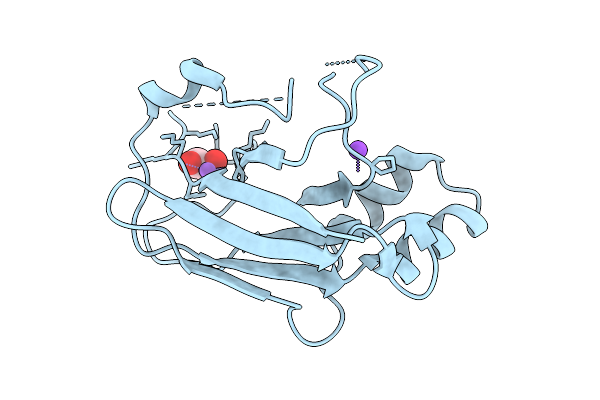
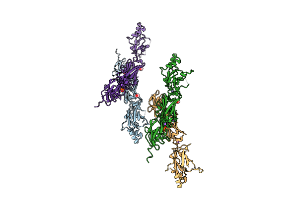
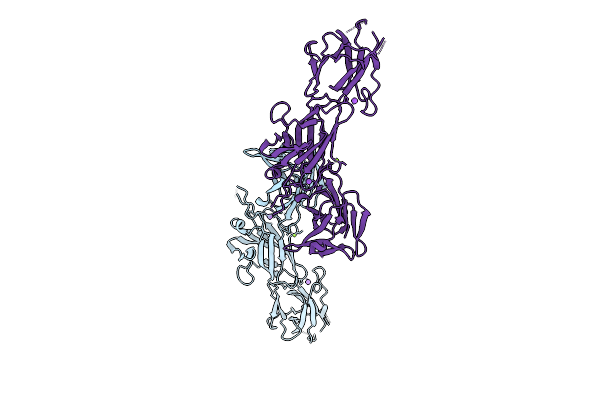
Organism: Homo sapiens, Jc polyomavirus
Method: X-RAY DIFFRACTION Resolution:3.17 Å Release Date: 2025-05-28 Classification: VIRAL PROTEIN Ligands: F6Z |
 |
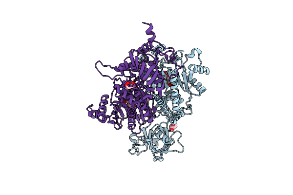
Respiratory Complex Peripheral Arm Of Ci, Close Form A, Focus-Refined Map Of Type I, Wild Type Mouse Under Thermoneutral Temperature
Organism: Mus musculus
Method: ELECTRON MICROSCOPY Release Date: 2024-10-30 Classification: ELECTRON TRANSPORT Ligands: 3PE, SF4, FES, FMN, UQ9, PC1, NDP, ZN, EHZ, CDL |
 |
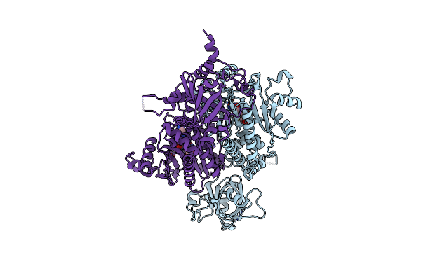
Respiratory Complex Peripheral Arm Of Ci, Close Form B, Focus-Refined Map Of Type I, Wild Type Mouse Under Thermoneutral Temperature
Organism: Mus musculus
Method: ELECTRON MICROSCOPY Release Date: 2024-10-30 Classification: ELECTRON TRANSPORT Ligands: 3PE, SF4, UQ9, FES, FMN, PC1, NDP, ZN, EHZ, CDL |
 |
Respiratory Complex Peripheral Arm Of Ci, Open Form A, Focus-Refined Map Of Type I, Wild Type Mouse Under Thermoneutral Temperature
Organism: Mus musculus
Method: ELECTRON MICROSCOPY Release Date: 2024-10-30 Classification: ELECTRON TRANSPORT Ligands: 3PE, SF4, FES, FMN, UQ9, PC1, NDP, ZN, EHZ, CDL |
 |
Respiratory Complex Peripheral Arm Of Ci, Close Form A, Focus-Refined Map Of Type Ia, Wild Type Mouse Under Cold Acclimation
Organism: Mus musculus
Method: ELECTRON MICROSCOPY Release Date: 2024-10-30 Classification: ELECTRON TRANSPORT Ligands: 3PE, SF4, PC1, UQ1, FES, FMN, UQ9, NDP, ZN, EHZ, CDL |