
Search Count: 885
 |
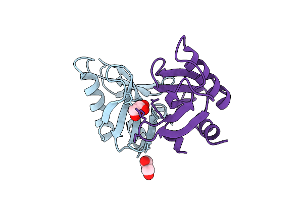
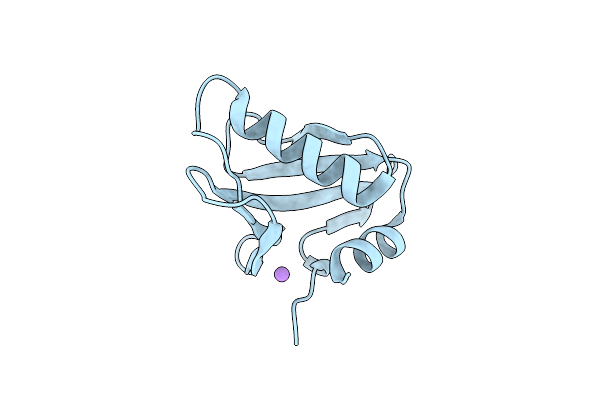
Crystal Structure Of The Chymotrypsin-Cleaved Iron-Free C-Lobe Of Bovine Lactoferrin At 2.82 Angstrom Resolution
Organism: Bos taurus
Method: X-RAY DIFFRACTION Release Date: 2025-12-31 Classification: METAL BINDING PROTEIN Ligands: EDO, GOL, ACT, NAG, SO4 |
 |
Organism: Microbacterium oxydans
Method: X-RAY DIFFRACTION Release Date: 2025-12-17 Classification: HYDROLASE Ligands: EDO |
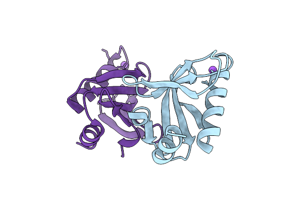 |
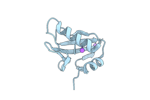
Crystal Structure Of Upf0235 Protein Pf1765 From Pyrococcus Furiosus Crystallized With Sodium Acetate At Ph 8
Organism: Pyrococcus furiosus dsm 3638
Method: X-RAY DIFFRACTION Resolution:1.30 Å Release Date: 2025-11-26 Classification: UNKNOWN FUNCTION |
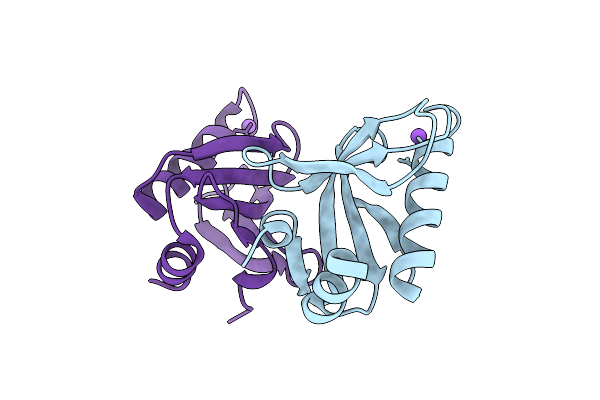 |
Crystal Structure Of Upf0235 Protein Pf1765 From Pyrococcus Furiosus At Ph 6
Organism: Pyrococcus furiosus dsm 3638
Method: X-RAY DIFFRACTION Resolution:1.13 Å Release Date: 2025-11-26 Classification: UNKNOWN FUNCTION |
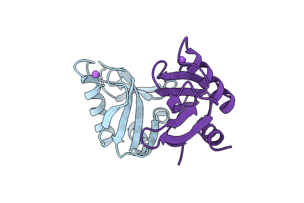 |
Crystal Structure Of Upf0235 Protein Pf1765 From Pyrococcus Furiosus At Ph 9
Organism: Pyrococcus furiosus dsm 3638
Method: X-RAY DIFFRACTION Resolution:1.30 Å Release Date: 2025-11-26 Classification: UNKNOWN FUNCTION |
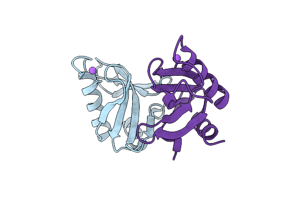 |
Crystal Structure Of Upf0235 Protein Pf1765 From Pyrococcus Furiosus Crystallized With Sodium Citrate At Ph 8.0
Organism: Pyrococcus furiosus dsm 3638
Method: X-RAY DIFFRACTION Resolution:1.45 Å Release Date: 2025-11-26 Classification: UNKNOWN FUNCTION |
 |
Crystal Structure Of Upf0235 Protein Pf1765 From Pyrococcus Furiosus Crystallized With Peg1500 And Glycerol At Ph 8.0
Organism: Pyrococcus furiosus dsm 3638
Method: X-RAY DIFFRACTION Resolution:1.11 Å Release Date: 2025-11-26 Classification: UNKNOWN FUNCTION Ligands: GOL, CL |
 |
Crystal Structure Of Upf0235 Protein Pf1765 From Pyrococcus Furiosus At Ph 4
Organism: Pyrococcus furiosus dsm 3638
Method: X-RAY DIFFRACTION Resolution:1.19 Å Release Date: 2025-11-26 Classification: UNKNOWN FUNCTION |
 |
Crystal Structure Of Upf0235 Protein Pf1765 From Pyrococcus Furiosus Crystallized With Cacl2 At Ph 5
Organism: Pyrococcus furiosus dsm 3638
Method: X-RAY DIFFRACTION Resolution:1.50 Å Release Date: 2025-11-26 Classification: UNKNOWN FUNCTION |
 |
Crystal Structure Of Upf0235 Protein Pf1765 From Pyrococcus Furiosus Crystallized With Cacl2 At Ph 6
Organism: Pyrococcus furiosus dsm 3638
Method: X-RAY DIFFRACTION Resolution:1.50 Å Release Date: 2025-11-26 Classification: UNKNOWN FUNCTION Ligands: CL, NA |
 |
Crystal Structure Of Upf0235 Protein Pf1765 From Pyrococcus Furiosus Crystallized With Cacl2 At Ph 7
Organism: Pyrococcus furiosus dsm 3638
Method: X-RAY DIFFRACTION Release Date: 2025-11-26 Classification: UNKNOWN FUNCTION |
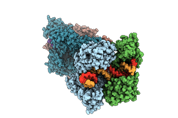 |
Crystal Structure Of Topoisomerase Iv From Klebsiella Pneumoniae In Complex With Dna And Bwc0977, A Dual-Targeting Broad-Spectrum Novel Bacterial Topoisomerase Inhibitor.
Organism: Klebsiella pneumoniae subsp. pneumoniae mgh 78578, Synthetic construct
Method: X-RAY DIFFRACTION Resolution:3.05 Å Release Date: 2025-11-12 Classification: ISOMERASE/DNA/ISOMERASE INHIBITOR Ligands: A1L5V |
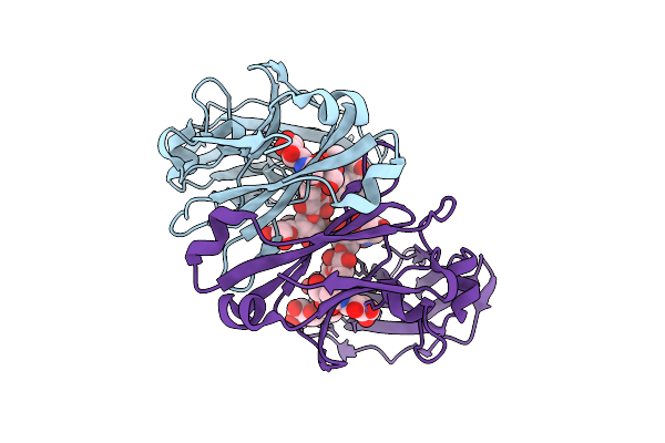 |
Crystal Structure Of Knob-In-Hole Immunoglobulin G1 Fc Heterodimer With P374A
Organism: Homo sapiens
Method: X-RAY DIFFRACTION Resolution:1.84 Å Release Date: 2025-11-12 Classification: IMMUNE SYSTEM |
 |
Crystal Structure Of Kirsten Rat Sarcoma G12C Complexed With Gmppnp And Covalently Bound To An Adduct Of {(2S)-4-[7-(8-Chloronaphthalen-1-Yl)-2-{[(2S)-1-Methylpyrrolidin-2-Yl]Methoxy}-5,6,7,8-Tetrahydropyrido[3,4-D]Pyrimidin-4-Yl]-1-[(2Z)-2-Fluoro-3-(Pyridin-2-Yl)Prop-2-Enoyl]Piperazin-2-Yl}Acetonitrile
Organism: Homo sapiens
Method: X-RAY DIFFRACTION Resolution:1.59 Å Release Date: 2025-11-05 Classification: HYDROLASE/HYDROLASE INHIBITOR Ligands: GNP, A1B7P, MG |
 |
Crystal Structure Of Kirsten Rat Sarcoma G12C Complexed With Gdp And Covalently Bound To An Adduct Of (2S)-1-{4-[(7P)-7-(8-Ethynyl-7-Fluoro-3-Hydroxynaphthalen-1-Yl)-8-Fluoro-2-{[(2R,4R,7As)-2-Fluorotetrahydro-1H-Pyrrolizin-7A(5H)-Yl]Methoxy}Pyrido[4,3-D]Pyrimidin-4-Yl]Piperazin-1-Yl}-2-Fluoro-3-(1,3-Thiazol-2-Yl)Propan-1-One
Organism: Homo sapiens
Method: X-RAY DIFFRACTION Resolution:1.50 Å Release Date: 2025-11-05 Classification: HYDROLASE/HYDROLASE INHIBITOR Ligands: GDP, A1B7Q, MG |
 |
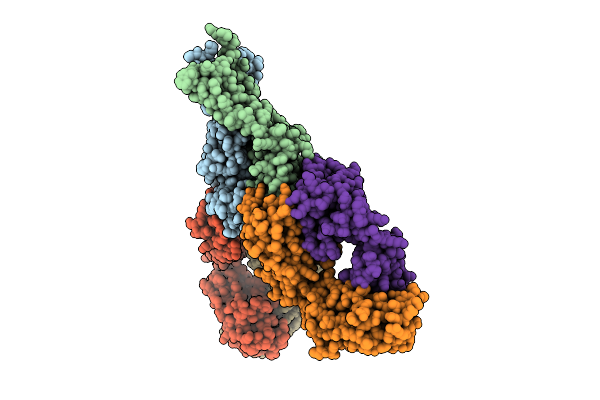
Organism: Sars-cov-2 pseudovirus, Homo sapiens
Method: ELECTRON MICROSCOPY Resolution:3.07 Å Release Date: 2025-10-29 Classification: MEMBRANE PROTEIN/IMMUNE SYSTEM/INHIBITOR Ligands: A1AE8 |
 |
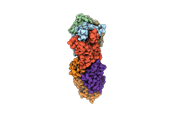
Organism: Severe acute respiratory syndrome coronavirus, Homo sapiens
Method: ELECTRON MICROSCOPY Release Date: 2025-10-29 Classification: MEMBRANE PROTEIN/IMMUNE SYSTEM/INHIBITOR |
 |
Organism: Middle east respiratory syndrome-related coronavirus, Homo sapiens
Method: ELECTRON MICROSCOPY Resolution:3.15 Å Release Date: 2025-10-29 Classification: MEMBRANE PROTEIN/IMMUNE SYSTEM/INHIBITOR |
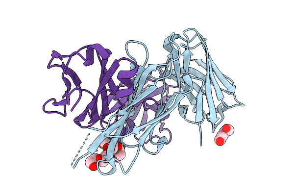 |
Organism: Homo sapiens
Method: X-RAY DIFFRACTION Resolution:2.60 Å Release Date: 2025-10-22 Classification: IMMUNE SYSTEM Ligands: PEG |
 |
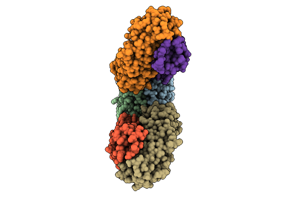
Cryo- Em Structure Of Large Subunit (Lsu) Of 75S Ribosome With P- Trna From Entamoeba Histolytica
Organism: Entamoeba histolytica hm-1:imss
Method: ELECTRON MICROSCOPY Release Date: 2025-10-15 Classification: RIBOSOME |