
Search Count: 25
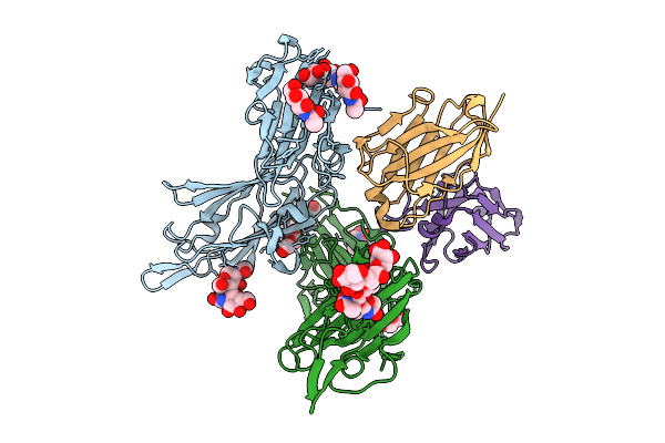 |
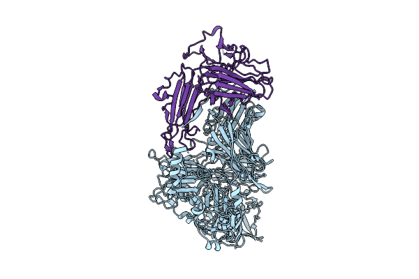
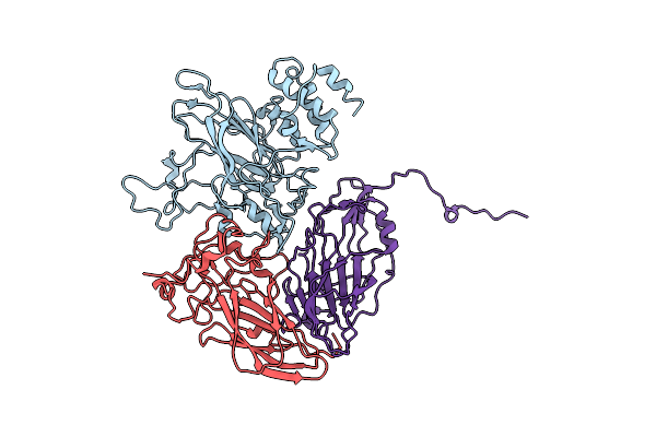
Organism: Plasmodium falciparum 3d7, Vicugna pacos
Method: X-RAY DIFFRACTION Release Date: 2025-07-30 Classification: PROTEIN BINDING Ligands: GOL, NAG, PEG |
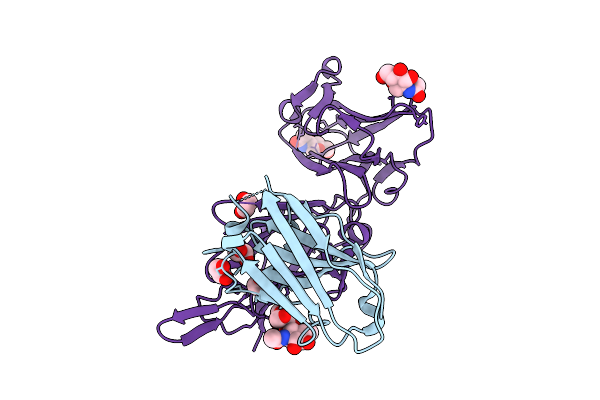 |
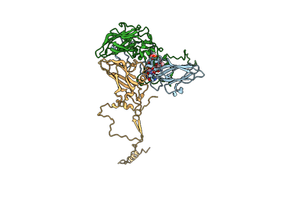
Organism: Vicugna pacos, Plasmodium falciparum 3d7
Method: X-RAY DIFFRACTION Release Date: 2025-07-30 Classification: PROTEIN BINDING Ligands: EDO, NAG |
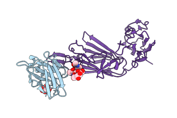 |
Organism: Vicugna pacos, Plasmodium falciparum 3d7
Method: X-RAY DIFFRACTION Release Date: 2025-07-30 Classification: PROTEIN BINDING Ligands: CIT |
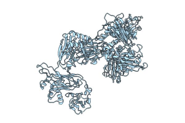 |
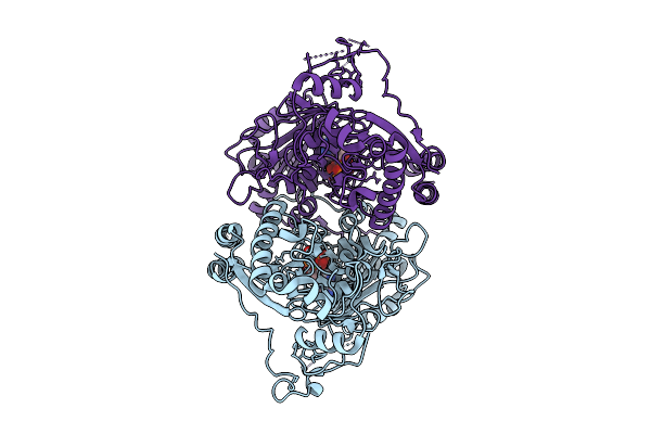
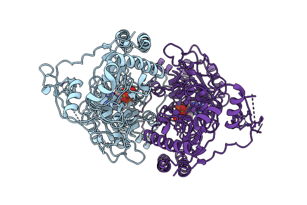
Organism: Plasmodium falciparum
Method: ELECTRON MICROSCOPY Release Date: 2025-07-30 Classification: PROTEIN BINDING |
 |
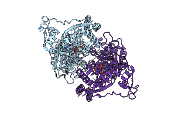
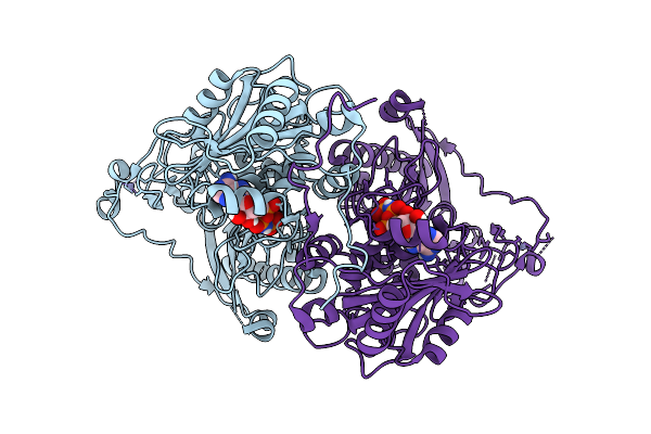
Organism: Plasmodium falciparum
Method: ELECTRON MICROSCOPY Release Date: 2025-07-30 Classification: PROTEIN BINDING |
 |
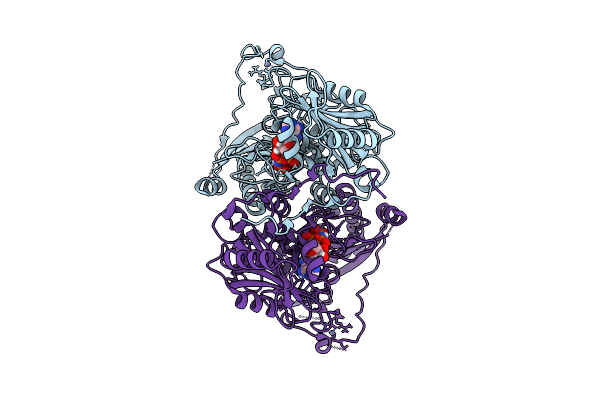
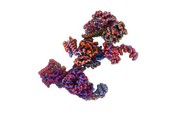
Organism: Homo sapiens
Method: ELECTRON MICROSCOPY Release Date: 2025-07-09 Classification: DNA BINDING PROTEIN Ligands: ZN, ANP, MG |
 |
Organism: Homo sapiens
Method: ELECTRON MICROSCOPY Release Date: 2025-07-09 Classification: DNA BINDING PROTEIN Ligands: ZN, ATP, MG |
 |
Organism: Homo sapiens
Method: ELECTRON MICROSCOPY Release Date: 2025-07-09 Classification: DNA BINDING PROTEIN Ligands: ZN, ANP, MG |
 |
Organism: Homo sapiens
Method: ELECTRON MICROSCOPY Release Date: 2025-07-09 Classification: DNA BINDING PROTEIN Ligands: ZN, ANP, MG |
 |
Organism: Homo sapiens
Method: ELECTRON MICROSCOPY Release Date: 2025-07-09 Classification: DNA BINDING PROTEIN Ligands: ZN, ANP, MG |
 |
Organism: Homo sapiens, Synthetic construct
Method: X-RAY DIFFRACTION Release Date: 2025-06-18 Classification: NUCLEAR PROTEIN |
 |
Organism: Homo sapiens, Synthetic construct
Method: X-RAY DIFFRACTION Release Date: 2025-06-11 Classification: DNA BINDING PROTEIN Ligands: MG |
 |
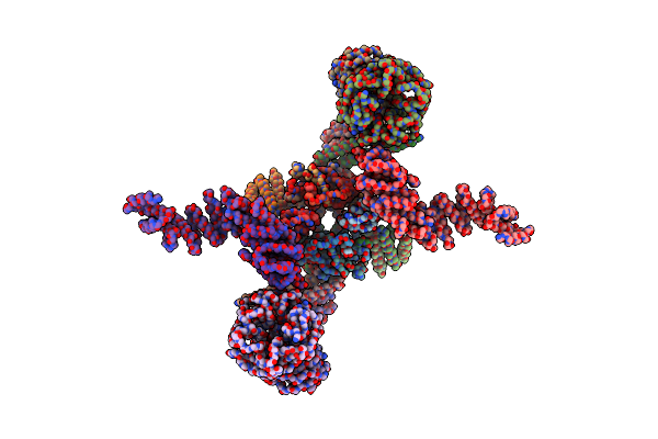
Structure Of The Dna-Bound Fancd2-Fanci Complex Containing Phosphomimetic Fanci
Organism: Gallus gallus, Synthetic construct
Method: ELECTRON MICROSCOPY Release Date: 2022-09-07 Classification: DNA BINDING PROTEIN |
 |
Organism: Gallus gallus, Homo sapiens, Synthetic construct
Method: ELECTRON MICROSCOPY Release Date: 2020-02-19 Classification: DNA BINDING PROTEIN |
 |
Organism: Gallus gallus
Method: ELECTRON MICROSCOPY Release Date: 2020-02-19 Classification: DNA BINDING PROTEIN |
 |
Organism: Gallus gallus
Method: ELECTRON MICROSCOPY Release Date: 2020-02-19 Classification: DNA BINDING PROTEIN |
 |
 |
 |
 |
Organism: Human parechovirus 1 (strain harris), Echovirus 22 (strain harris)
Method: X-RAY DIFFRACTION Resolution:3.09 Å Release Date: 2017-01-11 Classification: VIRUS |