
Search Count: 40
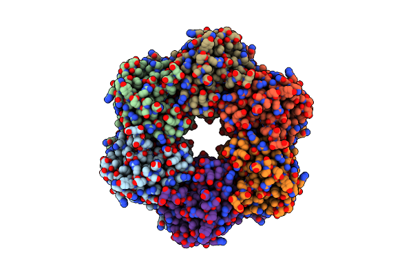 |
Organism: Synechococcus elongatus
Method: ELECTRON MICROSCOPY Release Date: 2025-05-14 Classification: CIRCADIAN CLOCK PROTEIN Ligands: ATP, MG |
 |
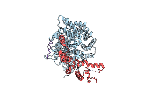
Organism: Synechococcus elongatus
Method: ELECTRON MICROSCOPY Release Date: 2025-05-14 Classification: CIRCADIAN CLOCK PROTEIN Ligands: ATP, MG |
 |
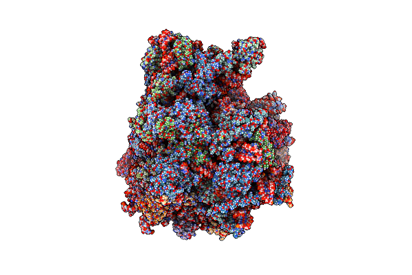
Organism: Synechococcus elongatus
Method: ELECTRON MICROSCOPY Release Date: 2025-05-14 Classification: CIRCADIAN CLOCK PROTEIN Ligands: ATP, MG |
 |
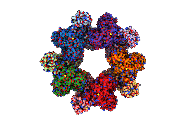
Organism: Influenza a virus, Homo sapiens
Method: ELECTRON MICROSCOPY Release Date: 2025-05-07 Classification: VIRAL PROTEIN Ligands: NAG |
 |
Organism: Influenza a virus
Method: X-RAY DIFFRACTION Release Date: 2025-05-07 Classification: VIRAL PROTEIN Ligands: NAG, GOL |
 |
Organism: Influenza a virus
Method: X-RAY DIFFRACTION Release Date: 2025-05-07 Classification: VIRAL PROTEIN Ligands: NAG |
 |
Organism: Homo sapiens, Respiratory syncytial virus a2
Method: X-RAY DIFFRACTION Resolution:1.74 Å Release Date: 2025-02-19 Classification: VIRAL PROTEIN/IMMUNE SYSTEM Ligands: SO4 |
 |
Organism: Homo sapiens, Respiratory syncytial virus a2
Method: X-RAY DIFFRACTION Resolution:2.50 Å Release Date: 2025-02-19 Classification: VIRAL PROTEIN/IMMUNE SYSTEM |
 |
Organism: Respiratory syncytial virus a2, Homo sapiens
Method: X-RAY DIFFRACTION Resolution:3.10 Å Release Date: 2025-02-19 Classification: VIRAL PROTEIN/IMMUNE SYSTEM |
 |
Organism: Homo sapiens
Method: ELECTRON MICROSCOPY Release Date: 2025-02-12 Classification: CELL CYCLE |
 |
Organism: Mus musculus, Human astrovirus 1
Method: ELECTRON MICROSCOPY Release Date: 2024-12-25 Classification: VIRAL PROTEIN/IMMUNE SYSTEM Ligands: NAG |
 |
Organism: Human astrovirus 2, Mus musculus
Method: X-RAY DIFFRACTION Release Date: 2024-12-25 Classification: VIRAL PROTEIN |
 |
Organism: Caenorhabditis elegans
Method: ELECTRON MICROSCOPY Release Date: 2024-09-11 Classification: RIBOSOME Ligands: SPD, 3HE |
 |
Organism: Caenorhabditis elegans
Method: ELECTRON MICROSCOPY Release Date: 2024-09-04 Classification: RIBOSOME Ligands: SPD, 3HE |
 |
Organism: Escherichia coli
Method: ELECTRON MICROSCOPY Release Date: 2024-04-24 Classification: RNA BINDING PROTEIN |
 |
Structure Of The Core Postfusion Porcine Endogenous Retrovirus Fusion Protein
Organism: Sus scrofa
Method: X-RAY DIFFRACTION Resolution:2.00 Å Release Date: 2022-01-12 Classification: VIRAL PROTEIN Ligands: CL, PEG |
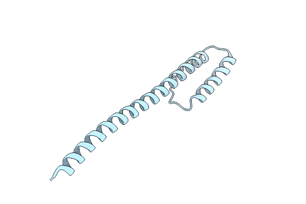 |
Structure Of An Influenza C Virus Hemagglutinin-Esterase-Fusion (Hef2) Intermediate
Organism: Influenza c virus (strain c/johannesburg/1/1966)
Method: X-RAY DIFFRACTION Resolution:2.40 Å Release Date: 2021-05-19 Classification: VIRAL PROTEIN Ligands: CL |
 |
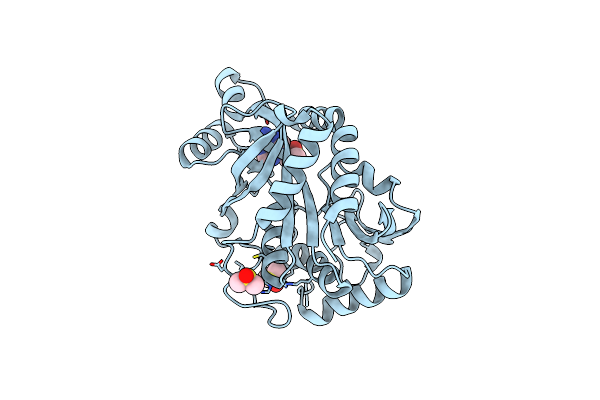
Crystal Structure Of Neisseria Meningitidis Clpp Protease Complex With Small Molecule Activator Acp1-06
Organism: Neisseria meningitidis
Method: X-RAY DIFFRACTION Resolution:1.64 Å Release Date: 2020-12-09 Classification: HYDROLASE Ligands: K, KHS |
 |
Crystal Structure Of Isoform 2 Of Purine Nucleoside Phosphorylase Complexed With Adenine
Organism: Schistosoma mansoni
Method: X-RAY DIFFRACTION Resolution:1.90 Å Release Date: 2017-10-11 Classification: TRANSFERASE Ligands: ADE, DMS |
 |
Crystal Structure Of Isoform 2 Of Purine Nucleoside Phosphorylase Complexed With Cytidine
Organism: Schistosoma mansoni
Method: X-RAY DIFFRACTION Resolution:2.10 Å Release Date: 2017-10-11 Classification: TRANSFERASE Ligands: CTN, SO4 |