
Search Count: 185
 |
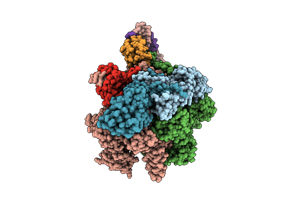
Cryoem Structure Of The T.Thermophilus Transcription Initiation Complex In The Presence Of Atp
Method: ELECTRON MICROSCOPY
Release Date: 2025-10-22 Classification: TRANSCRIPTION Ligands: MG, ZN, 1N7 |
 |
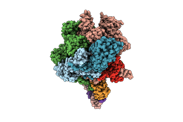
Cryoem Structure Of The T.Thermophilus Transcription Initiation Complex In The Presence Of Ap4A
Method: ELECTRON MICROSCOPY
Release Date: 2025-10-22 Classification: TRANSCRIPTION Ligands: ZN, MG, 1N7 |
 |
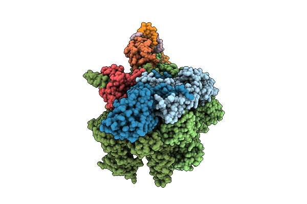
Cryoem Structure Of The T.Thermophilus Transcription Initiation Complex Bound To Atp-C, -1 Dc In The Template Dna Strand
Organism: Thermus thermophilus hb8
Method: ELECTRON MICROSCOPY Release Date: 2025-10-22 Classification: TRANSCRIPTION Ligands: MG, 1N7, ZN, C5P, ATP |
 |
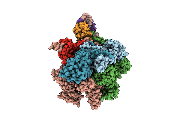
Cryoem Structure Of The T.Thermophilus Transcription Initiation Complex In The Presence Of Up4A
Method: ELECTRON MICROSCOPY
Release Date: 2025-10-22 Classification: TRANSCRIPTION Ligands: MG, 1N7, ZN |
 |
Method: ELECTRON MICROSCOPY
Release Date: 2025-10-22 Classification: TRANSCRIPTION Ligands: ZN, MG, 1N7 |
 |
Cryoem Structure Of The T.Thermophilus Transcription Initiation Complex Bound To Ap4A-C, -1 Da In The Template Dna Strand
Method: ELECTRON MICROSCOPY
Release Date: 2025-10-22 Classification: TRANSCRIPTION Ligands: MG, 1N7, ZN, C5P, B4P |
 |
Cryoem Structure Of The T.Thermophilus Transcription Initiation Complex Bound To Ap4A-C, -1 Dc In The Template Dna Strand
Method: ELECTRON MICROSCOPY
Release Date: 2025-10-22 Classification: TRANSCRIPTION Ligands: ZN, MG, C5P, B4P, 1N7 |
 |
Cryoem Structure Of The T.Thermophilus Transcription Initiation Complex In The Presence Of Gp4A, -1 Da In The Template Dna Strand
Method: ELECTRON MICROSCOPY
Release Date: 2025-10-22 Classification: TRANSCRIPTION Ligands: MG, 1N7, ZN |
 |
Cryoem Structure Of The T.Thermophilus Transcription Initiation Complex Bound To Gp4A-C-U And Ctp, -1 Dc In The Template Dna Strand
Method: ELECTRON MICROSCOPY
Release Date: 2025-10-22 Classification: TRANSCRIPTION Ligands: MG, 1N7, CTP, ZN |
 |
Cryoem Structure Of The T.Thermophilus Transcription Initiation Complex Bound To Gp4A-C, -1 Dc In The Template Dna Strand
Method: ELECTRON MICROSCOPY
Release Date: 2025-10-22 Classification: TRANSCRIPTION Ligands: ZN, MG, C5P, 1N7, A1IF9 |
 |
Cryoem Structure Of The T.Thermophilus Transcription Initiation Complex Bound To Up4A-C And Up4A, -1 Da In The Template Dna Strand
Method: ELECTRON MICROSCOPY
Release Date: 2025-10-22 Classification: TRANSCRIPTION Ligands: MG, 1N7, A1L89, ZN, C5P |
 |
Cryoem Structure Of The T.Thermophilus Transcription Initiation Complex Bound To Up4A-C And Utp, -1 Da In The Template Dna Strand
Organism: Thermus thermophilus hb8, Dna molecule
Method: ELECTRON MICROSCOPY Release Date: 2025-10-22 Classification: TRANSCRIPTION Ligands: MG, ZN, 1N7, C5P, UTP, A1L89 |
 |
Cryoem Structure Of T.Thermophilus Transcription Initiation Complex Bound To Gp4A-C, -1 Da In The Template Dna Strand
Method: ELECTRON MICROSCOPY
Release Date: 2025-10-22 Classification: TRANSCRIPTION Ligands: MG, 1N7, A1IF9, C5P, ZN |
 |
Cryoem Structure Of The T.Thermophilus Transcription Initiation Complex In The Presence Of Gp4A, -1 Dc In The Template Dna Strand
Method: ELECTRON MICROSCOPY
Release Date: 2025-10-22 Classification: TRANSCRIPTION Ligands: MG, 1N7, A1L9G, ZN |
 |
Crystal Structure Of The T.Thermophilus Transcription Initiation Complex Bound To Ap4G
Organism: Thermus thermophilus hb8, Synthetic construct
Method: X-RAY DIFFRACTION Release Date: 2025-10-22 Classification: TRANSCRIPTION Ligands: MG, A1IF9, TRS, ZN |
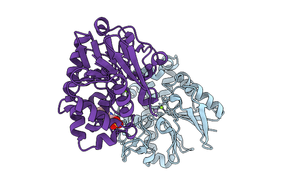 |
Organism: Escherichia coli k-12
Method: X-RAY DIFFRACTION Release Date: 2025-09-03 Classification: HYDROLASE Ligands: MN, MG, EPE |
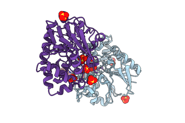 |
Organism: Escherichia coli k-12
Method: X-RAY DIFFRACTION Release Date: 2025-09-03 Classification: HYDROLASE Ligands: MN, SO4, EPE |
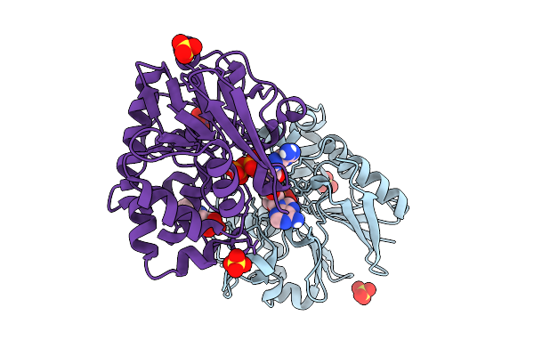 |
Organism: Escherichia coli k-12
Method: X-RAY DIFFRACTION Release Date: 2025-09-03 Classification: HYDROLASE Ligands: ADP, MN, MG, SO4, EPE |
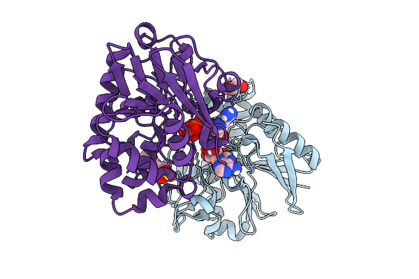 |
Organism: Escherichia coli k-12
Method: X-RAY DIFFRACTION Release Date: 2025-09-03 Classification: HYDROLASE Ligands: GOL, ADP, MN, EPE |
 |
Organism: Escherichia coli k-12
Method: X-RAY DIFFRACTION Release Date: 2025-09-03 Classification: HYDROLASE Ligands: GDP, SO4, MN, MG, EPE |