
Search Count: 18
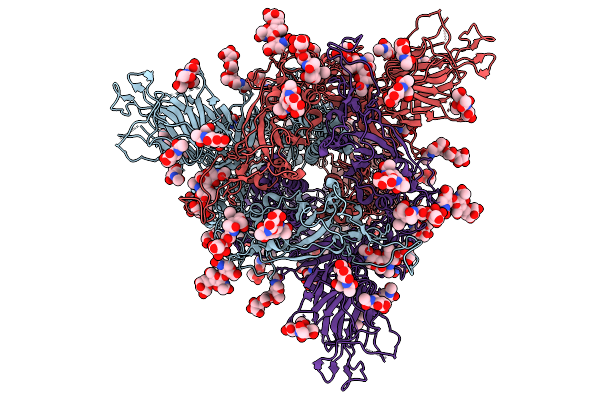 |
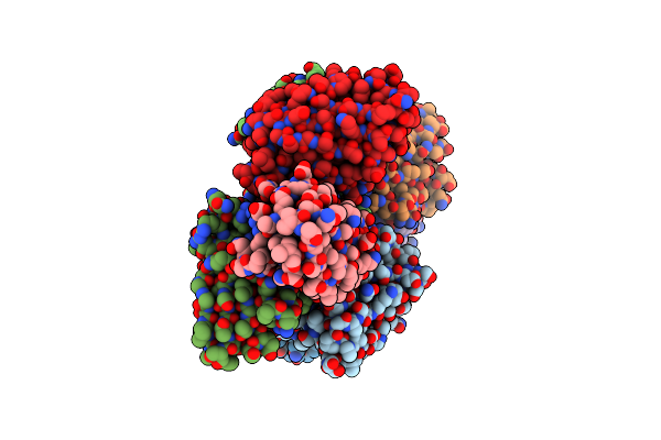
Organism: Sarbecovirus sp. rhgb07
Method: ELECTRON MICROSCOPY Release Date: 2025-09-24 Classification: VIRAL PROTEIN Ligands: NAG |
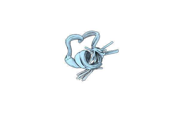 |
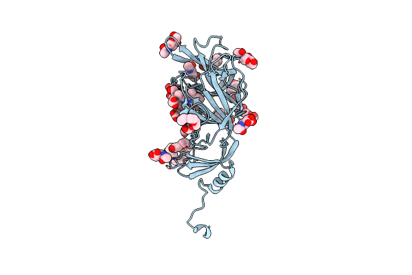
Structure Of Elevenin-Vc1 From Venom Of The Australian Cone Snail Conus Victoriae
|
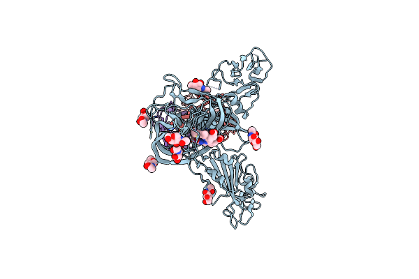 |
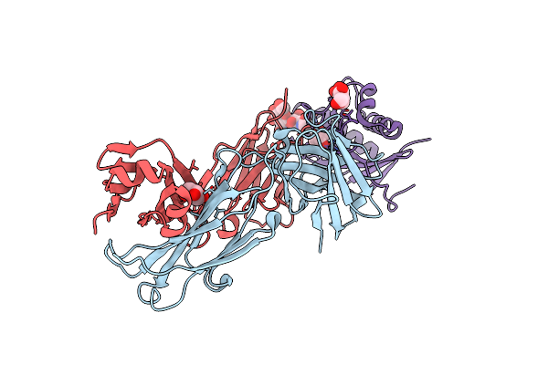
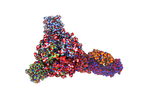
Cryoem Structure Of Sars-Cov-2 Spike Monomer In Complex With Neutralising Antibody P008_60
Organism: Severe acute respiratory syndrome coronavirus 2, Homo sapiens
Method: ELECTRON MICROSCOPY Release Date: 2022-08-17 Classification: VIRAL PROTEIN Ligands: NAG, 3Q9 |
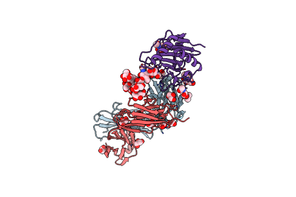 |
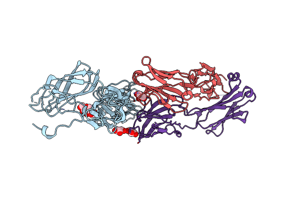
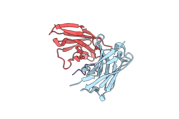
Machupo Virus Gp1 Glycoprotein In Complex With Fab Fragment Of Antibody Mac1
Organism: Mus musculus, Machupo mammarenavirus
Method: X-RAY DIFFRACTION Resolution:1.91 Å Release Date: 2022-02-09 Classification: IMMUNE SYSTEM Ligands: GOL, NAG |
 |
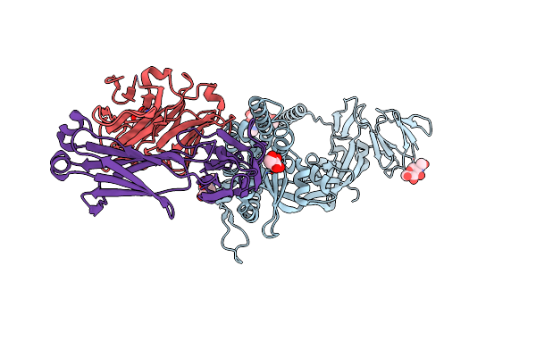
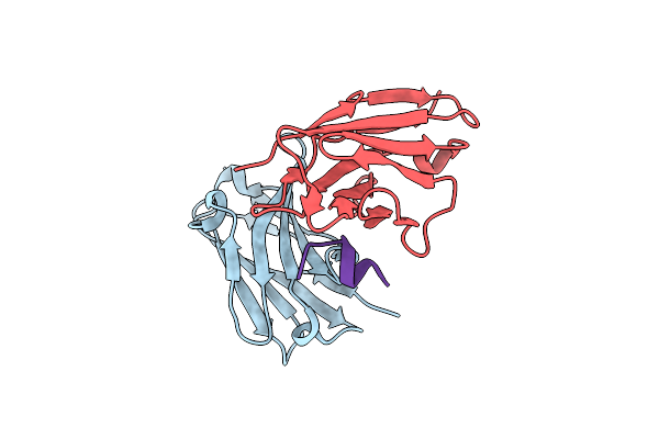
Organism: Mus musculus, Argentinian mammarenavirus
Method: X-RAY DIFFRACTION Resolution:2.50 Å Release Date: 2022-02-09 Classification: IMMUNE SYSTEM Ligands: GOL, NAG |
 |
Crystal Structure Of Sars-Cov-2 Spike Protein N-Terminal Domain In Complex With Biliverdin
Organism: Severe acute respiratory syndrome coronavirus 2
Method: X-RAY DIFFRACTION Resolution:1.82 Å Release Date: 2021-04-28 Classification: VIRAL PROTEIN Ligands: BLA, NAG, PG4, PEG, PGE, 1PE |
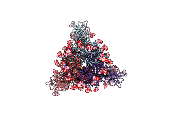 |
Trimeric Sars-Cov-2 Spike Ectodomain In Complex With Biliverdin (Closed Conformation)
Organism: Severe acute respiratory syndrome coronavirus 2
Method: ELECTRON MICROSCOPY Release Date: 2021-04-28 Classification: VIRAL PROTEIN Ligands: BLA, NAG |
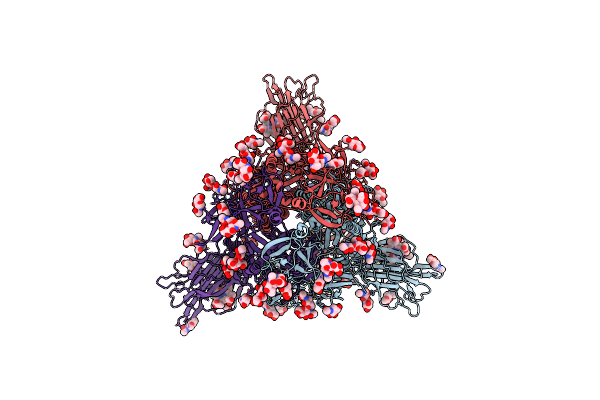 |
Trimeric Sars-Cov-2 Spike Ectodomain In Complex With Biliverdin (One Rbd Erect)
Organism: Severe acute respiratory syndrome coronavirus 2
Method: ELECTRON MICROSCOPY Release Date: 2021-04-28 Classification: VIRAL PROTEIN Ligands: BLA, NAG |
 |
Organism: Severe acute respiratory syndrome coronavirus 2, Homo sapiens
Method: ELECTRON MICROSCOPY Release Date: 2021-04-28 Classification: VIRAL PROTEIN Ligands: BLA, NAG |
 |
Organism: Myodes glareolus, Puumala orthohantavirus
Method: X-RAY DIFFRACTION Resolution:3.50 Å Release Date: 2020-12-30 Classification: VIRAL PROTEIN Ligands: GOL, NAG |
 |
Organism: Myodes glareolus, Puumala orthohantavirus
Method: ELECTRON MICROSCOPY Release Date: 2020-12-16 Classification: VIRUS LIKE PARTICLE Ligands: NAG |
 |
Organism: Puumala orthohantavirus
Method: ELECTRON MICROSCOPY Release Date: 2020-12-16 Classification: VIRUS LIKE PARTICLE Ligands: NAG |
 |
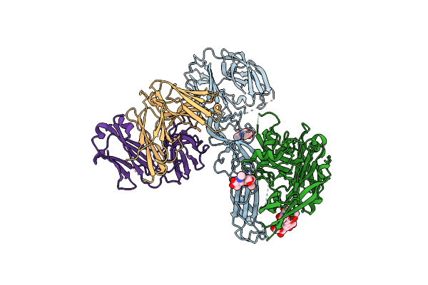
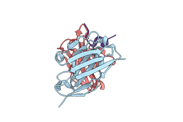
Crystal Structure Nipah Virus Fusion Glycoprotein In Complex With A Neutralising Fab Fragment
Organism: Nipah virus, Oryctolagus cuniculus
Method: X-RAY DIFFRACTION Resolution:3.20 Å Release Date: 2019-11-27 Classification: VIRAL PROTEIN Ligands: NAG |
 |
Crystal Structure Of Anti-Msp2 Fv Fragment (Mab4D11) In Complex With 3D7-Msp2 215-222
Organism: Mus musculus, Plasmodium falciparum 3d7
Method: X-RAY DIFFRACTION Resolution:2.20 Å Release Date: 2017-02-15 Classification: IMMUNE SYSTEM |
 |
Crystal Structure Of Anti-Msp2 Fv Fragment (Mab6D8)In Complex With Fc27-Msp2 14-30
Organism: Mus musculus, Plasmodium falciparum k1
Method: X-RAY DIFFRACTION Resolution:1.58 Å Release Date: 2015-06-03 Classification: IMMUNE SYSTEM |
 |
Crystal Structure Of Anti-Msp2 Fv Fragment (Mab6D8) In Complex With 3D7-Msp2 14-30
Organism: Mus musculus, Plasmodium falciparum 3d7
Method: X-RAY DIFFRACTION Resolution:1.35 Å Release Date: 2015-06-03 Classification: IMMUNE SYSTEM |
 |
Crystal Structure Of Anti-Msp2 Fv Fragment (Mab6D8)In Complex With Msp2 14-22
Organism: Mus musculus, Plasmodium falciparum k1
Method: X-RAY DIFFRACTION Resolution:1.21 Å Release Date: 2015-06-03 Classification: IMMUNE SYSTEM |
 |
Crystal Structure Of Anti-Msp2 Fv Fragment (Mab6D8)In Complex With Msp2 11-23
Organism: Mus musculus, Plasmodium falciparum
Method: X-RAY DIFFRACTION Resolution:1.70 Å Release Date: 2015-06-03 Classification: IMMUNE SYSTEM |