
Search Count: 23
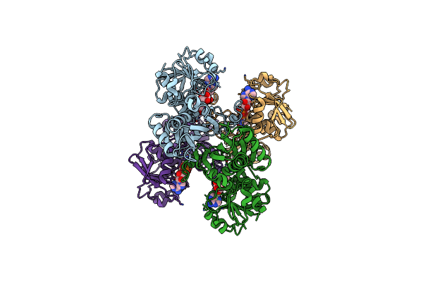 |
Crystal Structure Of Chlamydia Trachomatis Glyceraldehyde 3-Phosphate Dehydrogenase
Organism: Chlamydia trachomatis (strain d/uw-3/cx)
Method: X-RAY DIFFRACTION Resolution:1.50 Å Release Date: 2020-11-04 Classification: OXIDOREDUCTASE Ligands: NAD |
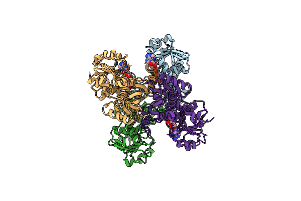 |
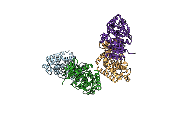
Crystal Structure Of Chlamydia Trachomatis Mixed (Apo/Holo) Glyceraldehyde 3-Phosphate Dehydrogenase
Organism: Chlamydia trachomatis (strain d/uw-3/cx)
Method: X-RAY DIFFRACTION Resolution:1.80 Å Release Date: 2020-11-04 Classification: OXIDOREDUCTASE Ligands: NAD |
 |
Crystal Structure Of The Lactate Dehydrogenase From Cryptosporidium Parvum Complexed With Cofactor (B-Nicotinamide Adenine Dinucleotide) And Inhibitor (Oxamic Acid)
Organism: Cryptosporidium parvum
Method: X-RAY DIFFRACTION Resolution:2.15 Å Release Date: 2014-12-17 Classification: Oxidoreductase/Oxidoreductase inhibitor Ligands: NAD, OXM, GOL |
 |
Crystal Structure Of The Lactate Dehydrogenase From Cryptosporidium Parvum Complexed With Substrate (Pyruvic Acid) And Cofactor Analog (3-Acetylpyridine Adenine Dinucleotide)
Organism: Cryptosporidium parvum
Method: X-RAY DIFFRACTION Resolution:2.00 Å Release Date: 2014-12-17 Classification: OXIDOREDUCTASE Ligands: A3D, PYR, GOL |
 |
Crystal Structure Of The Lactate Dehydrogenase From Cryptosporidium Parvum Complexed With Substrate (L-Lactic Acid) And Cofactor (B-Nicotinamide Adenine Dinucleotide)
Organism: Cryptosporidium parvum
Method: X-RAY DIFFRACTION Resolution:2.05 Å Release Date: 2014-12-17 Classification: OXIDOREDUCTASE Ligands: NAD, 2OP, GOL |
 |
Crystal Structure Of The Lactate Dehydrogenase From Cryptosporidium Parvum Complexed With Substrate (Pyruvic Acid) And Cofactor (B-Nicotinamide Adenine Dinucleotide)
Organism: Cryptosporidium parvum
Method: X-RAY DIFFRACTION Resolution:2.20 Å Release Date: 2014-12-17 Classification: OXIDOREDUCTASE Ligands: NAD, PYR, GOL |
 |
Organism: Cryptosporidium parvum
Method: X-RAY DIFFRACTION Resolution:2.10 Å Release Date: 2014-12-17 Classification: OXIDOREDUCTASE |
 |
Crystal Structure Of Glyceraldehyde-3-Phosphate Dehydrogenase From Streptococcus Agalactiae Nem316 At 2.46 Angstrom Resolution
Organism: Streptococcus agalactiae
Method: X-RAY DIFFRACTION Resolution:2.46 Å Release Date: 2014-10-15 Classification: OXIDOREDUCTASE Ligands: NAD, EDO |
 |
Organism: Cryptosporidium parvum
Method: X-RAY DIFFRACTION Resolution:2.50 Å Release Date: 2012-10-17 Classification: TRANSFERASE Ligands: SO4, GOL, ACT |
 |
Crystal Structure Of The Catalytic Domain Of Plasmodium Falciparum Arf Gtpase Activating Protein
Organism: Plasmodium falciparum 3d7
Method: X-RAY DIFFRACTION Resolution:2.40 Å Release Date: 2011-11-09 Classification: HYDROLASE ACTIVATOR Ligands: ZN, SO4 |
 |
Organism: Plasmodium falciparum
Method: X-RAY DIFFRACTION Resolution:2.50 Å Release Date: 2010-11-10 Classification: PROTEIN TRANSPORT Ligands: GDP, MG, SO4 |
 |
Crystal Structure Of T. Cruzi Dhfr-Ts With 3 High Affinity Dhfr Inhibitors: Dq1 Inhibitor Complex
Organism: Trypanosoma cruzi
Method: X-RAY DIFFRACTION Resolution:2.50 Å Release Date: 2010-06-09 Classification: Oxidoreductase,Transferase Ligands: DQ1, NAP, SO4, EDO |
 |
Structures Of Dihydrofolate Reductase-Thymidylate Synthase Of Trypanosoma Cruzi In The Folate-Free State And In Complex With Two Antifolate Drugs, Trimetrexate And Methotrexate
Organism: Trypanosoma cruzi
Method: X-RAY DIFFRACTION Resolution:3.00 Å Release Date: 2009-05-19 Classification: OXIDOREDUCTASE, TRANSFERASE Ligands: NAP, TMQ, SO4, EDO |
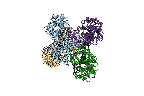 |
Crystal Structure Of Apo-Glyceraldehyde 3-Phosphate Dehydrogenase From Cryptosporidium Parvum
Organism: Cryptosporidium parvum
Method: X-RAY DIFFRACTION Resolution:2.20 Å Release Date: 2009-03-24 Classification: OXIDOREDUCTASE |
 |
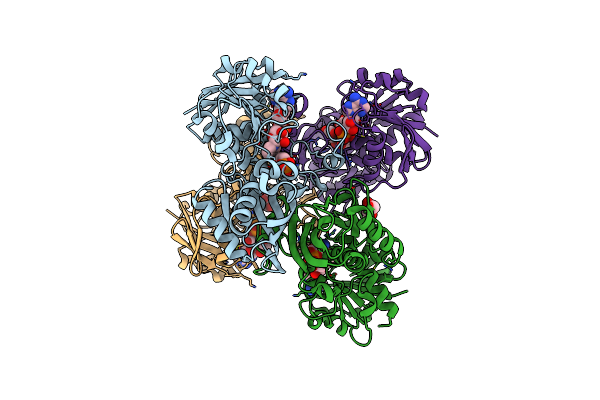
Crystal Structure Of Holo-Glyceraldehyde 3-Phosphate Dehydrogenase From Cryptosporidium Parvum
Organism: Cryptosporidium parvum
Method: X-RAY DIFFRACTION Resolution:2.00 Å Release Date: 2009-03-24 Classification: OXIDOREDUCTASE Ligands: NAD |
 |
Crystal Structure Of C153S Mutant Glyceraldehyde 3-Phosphate Dehydrogenase From Cryptosporidium Parvum
Organism: Cryptosporidium parvum
Method: X-RAY DIFFRACTION Resolution:2.00 Å Release Date: 2009-03-24 Classification: OXIDOREDUCTASE Ligands: G3H, NAD, GOL, MG |
 |
Organism: Trypanosoma cruzi
Method: X-RAY DIFFRACTION Resolution:3.30 Å Release Date: 2009-01-06 Classification: OXIDOREDUCTASE, TRANSFERASE Ligands: SO4, CL, NAP, MTX, UMP, EDO |
 |
Organism: Trypanosoma cruzi
Method: X-RAY DIFFRACTION Resolution:3.00 Å Release Date: 2009-01-06 Classification: OXIDOREDUCTASE, TRANSFERASE Ligands: SO4, NAP, TMQ, EDO |
 |
Crystal Structure Of Trypanosoma Cruzi Dihydrofolate Reductase-Thymidylate Synthase
Organism: Trypanosoma cruzi
Method: X-RAY DIFFRACTION Resolution:2.40 Å Release Date: 2008-04-08 Classification: OXIDOREDUCTASE, TRANSFERASE Ligands: NAP, DU |
 |
Crystal Structure Of The Complex Of Human Lactoferrin N-Lobe And Lactoferrin-Binding Domain Of Pneumococcal Surface Protein A
Organism: Homo sapiens, Streptococcus pneumoniae
Method: X-RAY DIFFRACTION Resolution:2.91 Å Release Date: 2007-06-19 Classification: METAL TRANSPORT, HYDROLASE Ligands: NAG, CO3, FE, SO4, ZN |