
Search Count: 1,260
 |
Organism: Homo sapiens, Mus musculus
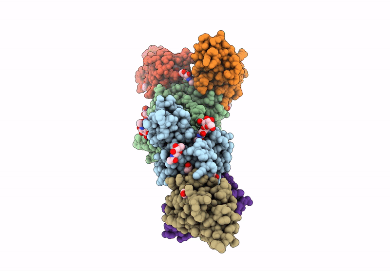
Method: ELECTRON MICROSCOPY Release Date: 2025-07-23 Classification: IMMUNE SYSTEM |
 |
Organism: Lotus japonicus
Method: X-RAY DIFFRACTION Release Date: 2025-07-16 Classification: PLANT PROTEIN Ligands: NAG, ACT, GOL |
 |
Organism: Medicago truncatula
Method: X-RAY DIFFRACTION Release Date: 2025-07-16 Classification: PLANT PROTEIN Ligands: NAG, SO4, EDO |
 |
Organism: Medicago truncatula
Method: X-RAY DIFFRACTION Release Date: 2025-07-16 Classification: PLANT PROTEIN Ligands: NAG |
 |
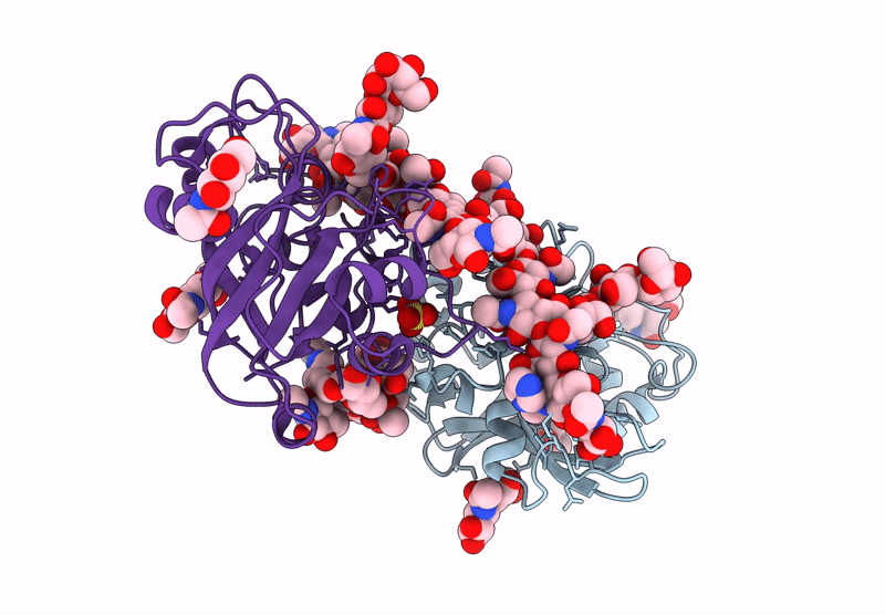
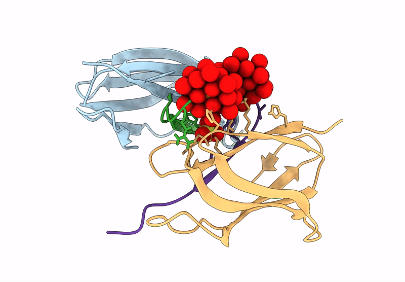
Crystal Structure Of Lotus Japonicus Chip13 Extracellular Domain In Complex With A Nanobody
Organism: Lotus japonicus, Lama glama
Method: X-RAY DIFFRACTION Release Date: 2025-07-16 Classification: PLANT PROTEIN Ligands: NAG |
 |
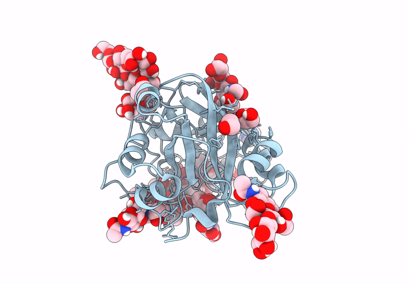
Crystal Structure Of Lotus Japonicus Chip13 Extracellular Domain In Complex With Chitooctaose
Organism: Lotus japonicus
Method: X-RAY DIFFRACTION Release Date: 2025-07-16 Classification: PLANT PROTEIN Ligands: GOL, NAG |
 |
Organism: Lotus japonicus
Method: X-RAY DIFFRACTION Release Date: 2025-07-16 Classification: PLANT PROTEIN |
 |
Organism: Lotus japonicus, Lama glama
Method: X-RAY DIFFRACTION Release Date: 2025-07-16 Classification: PLANT PROTEIN Ligands: NAG, EDO |
 |
Organism: Lotus japonicus
Method: X-RAY DIFFRACTION Release Date: 2025-07-16 Classification: PLANT PROTEIN Ligands: ACT, IMD, GOL, SO4, NAG |
 |
Crystal Structure Of Lotus Japonicus Chip13 Extracellular Domain In Complex With Chitooctaose
Organism: Lotus japonicus
Method: X-RAY DIFFRACTION Release Date: 2025-07-16 Classification: PLANT PROTEIN Ligands: IMD, EDO |
 |
Organism: Lotus japonicus
Method: X-RAY DIFFRACTION Release Date: 2025-07-16 Classification: PLANT PROTEIN Ligands: NAG, EDO, MAN |
 |
Crystal Structure Of Homolog Of Dihydroxyacid Dehydratase(Astd) From Aspergillus Terreus
Organism: Aspergillus terreus
Method: X-RAY DIFFRACTION Release Date: 2025-07-09 Classification: LYASE Ligands: PEG, EDO |
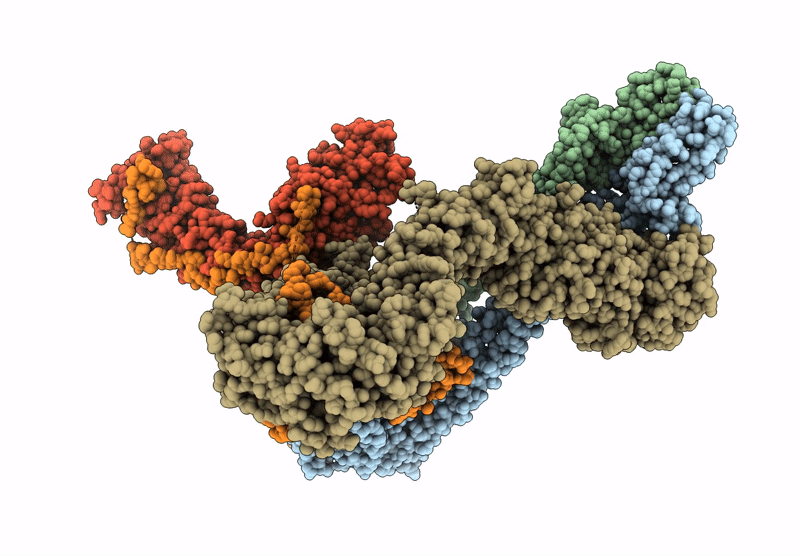 |
Organism: Homo sapiens, Escherichia coli (strain k12)
Method: ELECTRON MICROSCOPY Release Date: 2025-07-02 Classification: DNA BINDING PROTEIN |
 |
Organism: Streptococcus pyogenes
Method: X-RAY DIFFRACTION Release Date: 2025-06-25 Classification: PEPTIDE BINDING PROTEIN Ligands: TEW, SO4 |
 |
Organism: Plasmodium vivax
Method: X-RAY DIFFRACTION Resolution:2.06 Å Release Date: 2025-06-04 Classification: LIGASE Ligands: SO4, EDO |
 |
Plasmodium Vivax Aspartyl-Trna Synthetase In Complex With Aspartyl-Adenylate (Asp-Amp) Complex.
Organism: Plasmodium vivax
Method: X-RAY DIFFRACTION Resolution:2.14 Å Release Date: 2025-06-04 Classification: LIGASE Ligands: AMO, GOL, ACT |
 |
Plasmodium Vivax Aspartyl-Trna Synthetase In Complex With Aspartyl Sulfamoyl Adenosine (Asp-Ams) Complex
Organism: Plasmodium vivax
Method: X-RAY DIFFRACTION Resolution:1.84 Å Release Date: 2025-06-04 Classification: LIGASE Ligands: DSZ, MG |
 |
Structural Studies Of Reaction Hijacking Inhibition Of A Malaria Parasite Aspartyl-Trna Synthetase.
Organism: Plasmodium vivax
Method: X-RAY DIFFRACTION Release Date: 2025-06-04 Classification: LIGASE Ligands: A1B0L, GOL |
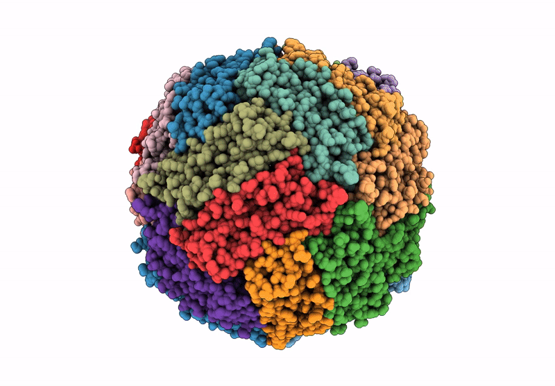 |
C-Terminus Truncated (Last Two Residues) Mutant Of Human Light Chain Ferritin Reacted With Ferrous Salt(3 Fe2+ Per Ferritin Subunit) . Reconstruction Of Particles With One Nanoparticle
Organism: Homo sapiens
Method: ELECTRON MICROSCOPY Release Date: 2025-05-14 Classification: METAL BINDING PROTEIN |
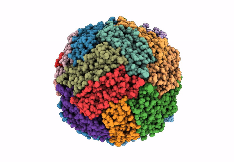 |
Human Light Chain Ferritin Reacted With Iron (3 Fe2+ To Ferritin Monomer Ratio). Reconstruction Of Particles With One Nanoparticle
Organism: Homo sapiens
Method: ELECTRON MICROSCOPY Release Date: 2025-05-14 Classification: METAL BINDING PROTEIN |