
Search Count: 37
 |
Organism: Saccharomyces cerevisiae
Method: ELECTRON MICROSCOPY Release Date: 2024-07-24 Classification: LIPID BINDING PROTEIN |
 |
Organism: Saccharomyces cerevisiae
Method: ELECTRON MICROSCOPY Release Date: 2024-07-24 Classification: LIPID BINDING PROTEIN |
 |
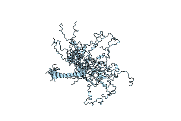
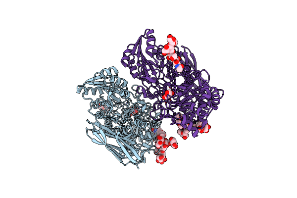
Helical Reconstruction Of Yeast Eisosome Protein Pil1 Bound To Membrane Composed Of Lipid Mixture -Pip2/+Sterol (Dopc, Dope, Dops, Cholesterol 30:20:20:30)
Organism: Saccharomyces cerevisiae
Method: ELECTRON MICROSCOPY Release Date: 2024-07-24 Classification: LIPID BINDING PROTEIN |
 |
Helical Reconstruction Of Yeast Eisosome Protein Pil1 Bound To Membrane Composed Of Lipid Mixture +Pip2/-Sterol (Dopc, Dope, Dops, Pi(4,5)P2 50:20:20:10)
Organism: Saccharomyces cerevisiae
Method: ELECTRON MICROSCOPY Release Date: 2024-07-24 Classification: LIPID BINDING PROTEIN Ligands: I3P |
 |
Helical Reconstruction Of Yeast Eisosome Protein Pil1 Bound To Membrane Composed Of Lipid Mixture +Pip2/+Sterol (Dopc, Dope, Dops, Cholesterol, Pi(4,5)P2 35:20:20:15:10)
Organism: Saccharomyces cerevisiae
Method: ELECTRON MICROSCOPY Release Date: 2024-07-24 Classification: LIPID BINDING PROTEIN Ligands: I3P, P5S |
 |
Compact State - Pil1 In Native Eisosome Lattice Bound To Plasma Membrane Microdomain
Organism: Saccharomyces cerevisiae
Method: ELECTRON MICROSCOPY Release Date: 2024-07-24 Classification: LIPID BINDING PROTEIN |
 |
Compact State - Pil1 Dimer With Lipid Headgroups Fitted In Native Eisosome Lattice Bound To Plasma Membrane Microdomain
Organism: Saccharomyces cerevisiae
Method: ELECTRON MICROSCOPY Release Date: 2024-07-24 Classification: LIPID BINDING PROTEIN Ligands: I3P, SEP |
 |
Stretched State - Pil1 In Native Eisosome Lattice Bound To Plasma Membrane Microdomain
Organism: Saccharomyces cerevisiae
Method: ELECTRON MICROSCOPY Release Date: 2024-07-24 Classification: LIPID BINDING PROTEIN |
 |
Organism: Phanerochaete chrysosporium
Method: X-RAY DIFFRACTION Resolution:2.54 Å Release Date: 2022-02-09 Classification: HYDROLASE Ligands: NAG, GOL |
 |
Organism: Phanerochaete chrysosporium
Method: X-RAY DIFFRACTION Resolution:3.08 Å Release Date: 2022-02-09 Classification: HYDROLASE Ligands: 1PE, PEG, EDO, NAG, XYP |
 |
N-Terminal Domain Of Dynein Intermediate Chain From Chaetomium Thermophilum
Organism: Chaetomium thermophilum (strain dsm 1495 / cbs 144.50 / imi 039719)
Method: SOLUTION NMR Release Date: 2020-08-19 Classification: MOTOR PROTEIN |
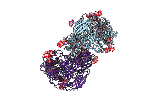 |
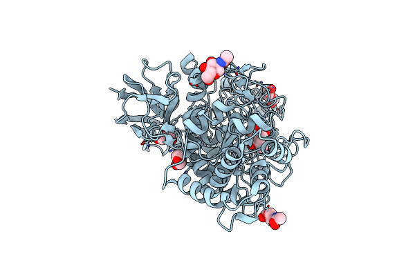
Structural Studies Of A Glycoside Hydrolase Family 3 Beta-Glucosidase From The Model Fungus Neurospora Crassa
Organism: Neurospora crassa or74a
Method: X-RAY DIFFRACTION Resolution:2.25 Å Release Date: 2018-03-21 Classification: HYDROLASE Ligands: NAG |
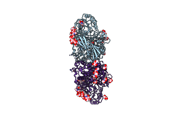 |
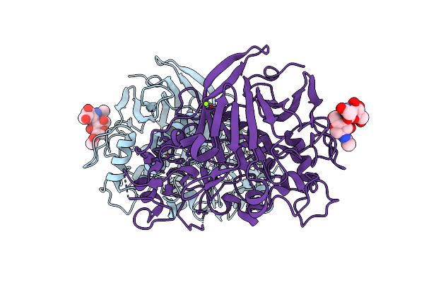
The Structure Of Hypocrea Jecorina Beta-Xylosidase Xyl3A (Bxl1) In Complex With 4-Thioxylobiose
Organism: Hypocrea jecorina
Method: X-RAY DIFFRACTION Resolution:2.10 Å Release Date: 2016-09-21 Classification: HYDROLASE Ligands: NAG, ZN |
 |
Organism: Trichoderma reesei
Method: X-RAY DIFFRACTION Resolution:1.80 Å Release Date: 2016-08-10 Classification: HYDROLASE Ligands: NAG, ZN, TRS, GOL, ACT |
 |
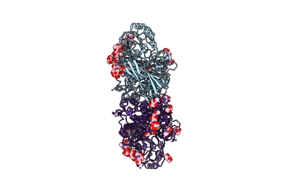
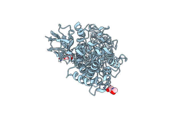
Crystal Structure Of Beta-D-Glucoside Glucohydrolase From Trichoderma Reesei
Organism: Trichoderma reesei
Method: X-RAY DIFFRACTION Resolution:2.48 Å Release Date: 2012-12-19 Classification: HYDROLASE Ligands: NAG, BGC |
 |
Crystal Structure Of A Glycoside Hydrolase Family 3 Beta-Glucosidase, Bgl1 From Hypocrea Jecorina At 2.1A Resolution.
Organism: Hypocrea jecorina
Method: X-RAY DIFFRACTION Resolution:2.10 Å Release Date: 2012-12-12 Classification: HYDROLASE Ligands: NAG, BGC, GOL, PEG |
 |
Crystal Structure Of A Glycoside Hydrolase Family 3 Beta-Glucosidase, Bgl1 From Hypocrea Jecorina At 2.1A Resolution.
Organism: Hypocrea jecorina
Method: X-RAY DIFFRACTION Resolution:2.10 Å Release Date: 2012-12-12 Classification: HYDROLASE Ligands: NAG, GOL |
 |
Organism: Heterobasidion annosum
Method: X-RAY DIFFRACTION Resolution:1.90 Å Release Date: 2012-04-25 Classification: HYDROLASE Ligands: MG |
 |
Elucidation Of The Substrate Specificity And Protein Structure Of Abfb, A Family 51 Alpha-L-Arabinofuranosidase From Bifidobacterium Longum.
Organism: Bifidobacterium longum
Method: X-RAY DIFFRACTION Resolution:2.50 Å Release Date: 2011-12-28 Classification: HYDROLASE |
 |
Organism: Heterobasidion annosum
Method: X-RAY DIFFRACTION Resolution:1.70 Å Release Date: 2011-10-12 Classification: HYDROLASE Ligands: EPE, XYS, MG |