
Search Count: 567
 |
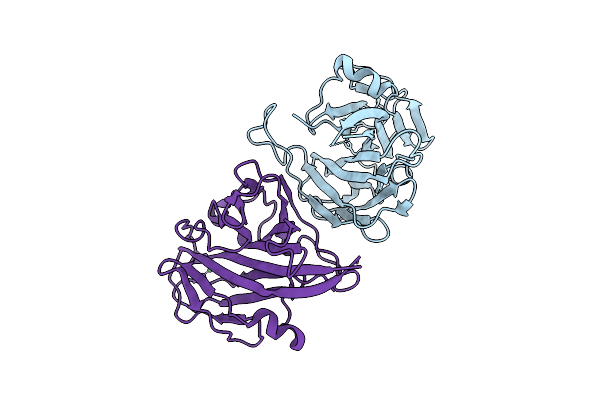
Organism: Neisseria gonorrhoeae fa 1090
Method: X-RAY DIFFRACTION Release Date: 2025-12-17 Classification: METAL BINDING PROTEIN |
 |
Organism: Neisseria gonorrhoeae fa 1090
Method: X-RAY DIFFRACTION Release Date: 2025-12-17 Classification: METAL BINDING PROTEIN |
 |
Organism: Neisseria gonorrhoeae fa 1090
Method: X-RAY DIFFRACTION Release Date: 2025-12-17 Classification: METAL BINDING PROTEIN |
 |
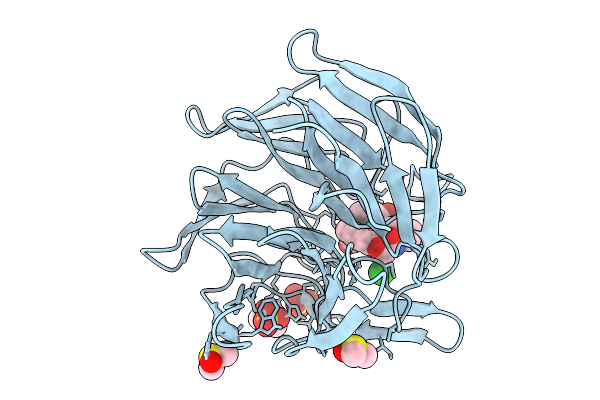
Organism: Neisseria gonorrhoeae fa 1090
Method: X-RAY DIFFRACTION Release Date: 2025-12-17 Classification: METAL BINDING PROTEIN Ligands: ZN |
 |
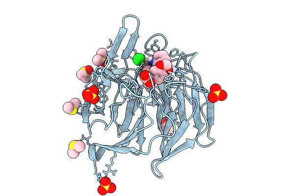
Organism: Neisseria gonorrhoeae fa 1090
Method: X-RAY DIFFRACTION Release Date: 2025-12-17 Classification: METAL BINDING PROTEIN Ligands: CU |
 |
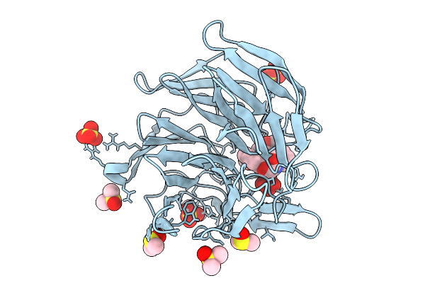
Organism: Neisseria gonorrhoeae fa 1090
Method: X-RAY DIFFRACTION Release Date: 2025-12-17 Classification: METAL BINDING PROTEIN Ligands: CO |
 |
Organism: Neisseria gonorrhoeae fa 1090
Method: X-RAY DIFFRACTION Release Date: 2025-12-17 Classification: METAL BINDING PROTEIN Ligands: NI |
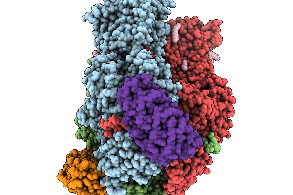 |
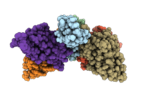
Structure Of The Double Cys-Substituted Cross-Linked Acrb Variant S562C_T837C
Organism: Escherichia coli k-12, Synthetic construct
Method: X-RAY DIFFRACTION Release Date: 2025-11-19 Classification: TRANSPORT PROTEIN Ligands: LMT, D10, GOL, D12, HEX, C14, OCT, DD9, SO4 |
 |
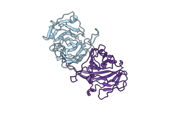
Organism: Homo sapiens, Bungarus multicinctus
Method: X-RAY DIFFRACTION Release Date: 2025-11-19 Classification: TOXIN Ligands: EDO |
 |
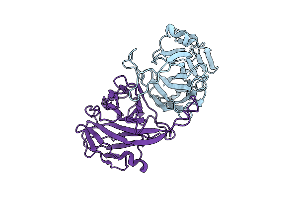
Organism: Homo sapiens, Naja kaouthia
Method: X-RAY DIFFRACTION Release Date: 2025-11-19 Classification: TOXIN Ligands: GOL, CL, K, NA, SO4 |
 |
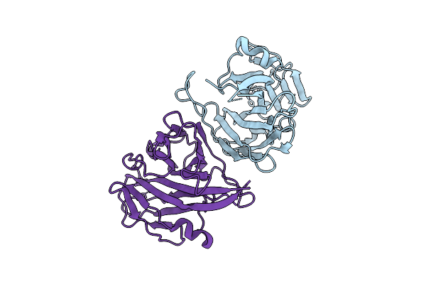
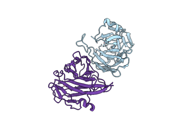
Organism: Homo sapiens, Naja kaouthia
Method: X-RAY DIFFRACTION Release Date: 2025-11-19 Classification: TOXIN Ligands: CL, GOL, TRS, SO4, NA |
 |
Organism: Escherichia coli k-12, Synthetic construct
Method: X-RAY DIFFRACTION Release Date: 2025-11-12 Classification: TRANSPORT PROTEIN Ligands: LMT, OCT, C14, EDO, GOL, D12, HEX, XE9, D10, DDR, DDQ, SO4, LPX |
 |
Organism: Gallus gallus
Method: X-RAY DIFFRACTION Release Date: 2025-10-29 Classification: TRANSPORT PROTEIN Ligands: HEM, CA |
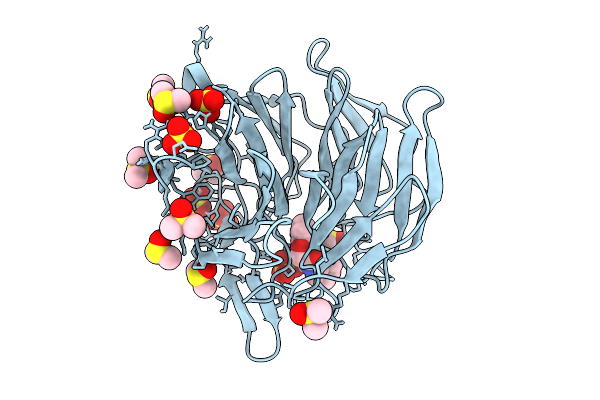 |
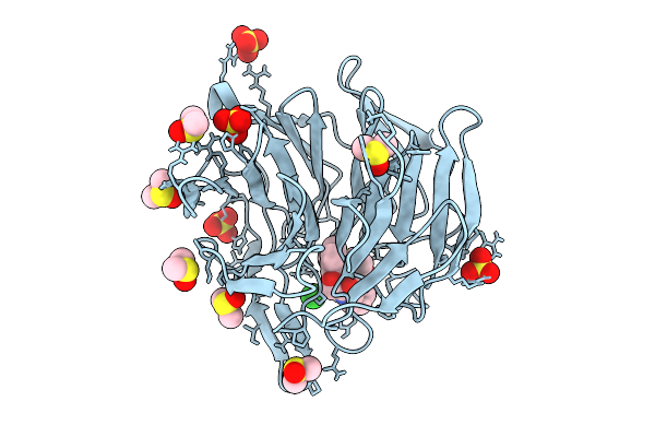
Crystal Structure Of The Keap1 Kelch Domain In Complex With The Xchem Fragment Z19735904 At 1.14 Angstrom Resolution.
Organism: Mus musculus
Method: X-RAY DIFFRACTION Release Date: 2025-09-03 Classification: PEPTIDE BINDING PROTEIN Ligands: B0A, SO4, DMS |
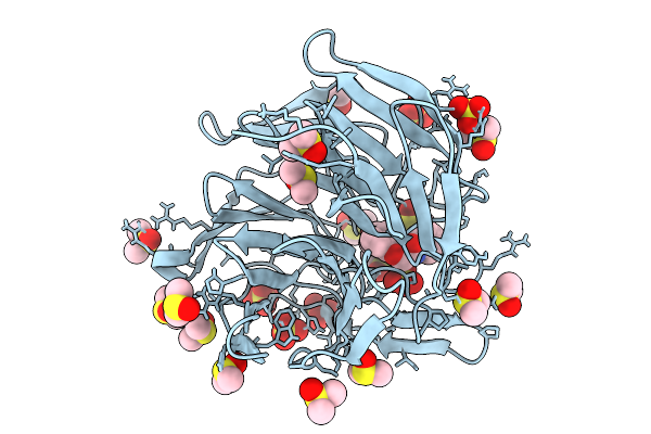 |
Crystal Structure Of The Keap1 Kelch Domain In Complex With The Small Molecule Ucab#827 At 1.40 Angstrom Resolution
Organism: Mus musculus
Method: X-RAY DIFFRACTION Release Date: 2025-09-03 Classification: PEPTIDE BINDING PROTEIN Ligands: A1IX2, CL, SO4, DMS |
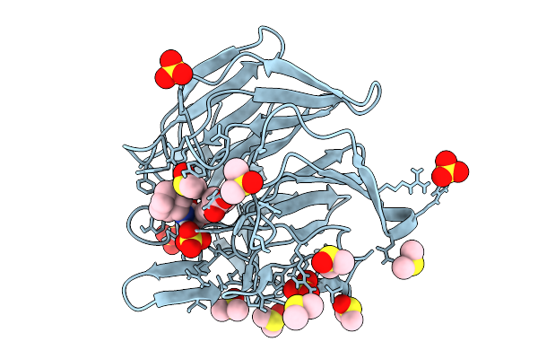 |
Crystal Structure Of The Keap1 Kelch Domain In Complex With The Small Molecule Ucab#909 At 1.61 Angstrom Resolution
Organism: Mus musculus
Method: X-RAY DIFFRACTION Release Date: 2025-09-03 Classification: PEPTIDE BINDING PROTEIN Ligands: A1IX3, SO4, DMS, CL |
 |
Crystal Structure Of The Keap1 Kelch Domain In Complex With The Small Molecule Ucab#985 At 1.65 Angstrom Resolution
Organism: Mus musculus
Method: X-RAY DIFFRACTION Release Date: 2025-09-03 Classification: PEPTIDE BINDING PROTEIN Ligands: A1IX4, SO4, DMS, CL |
 |
Crystal Structure Of The Keap1 Kelch Domain In Complex With The Small Molecule Ucab#1004 At 1.40 Angstrom Resolution
Organism: Mus musculus
Method: X-RAY DIFFRACTION Release Date: 2025-09-03 Classification: PEPTIDE BINDING PROTEIN Ligands: A1IXY, SO4, CL, DMS |
 |
Crystal Structure Of The Keap1 Kelch Domain In Complex With The Small Molecule Ucab#1010 At 1.50 Angstrom Resolution
Organism: Mus musculus
Method: X-RAY DIFFRACTION Release Date: 2025-09-03 Classification: PEPTIDE BINDING PROTEIN Ligands: A1IXZ, SO4, CL, DMS |
 |
Crystal Structure Of The Keap1 Kelch Domain In Complex With The Small Molecule Ucab#1032 At 1.61 Angstrom Resolution
Organism: Mus musculus
Method: X-RAY DIFFRACTION Release Date: 2025-09-03 Classification: PEPTIDE BINDING PROTEIN Ligands: A1IX0, SO4, DMS |