
Search Count: 36
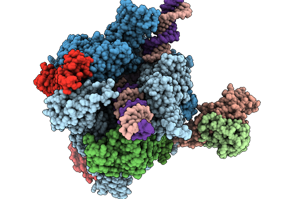 |
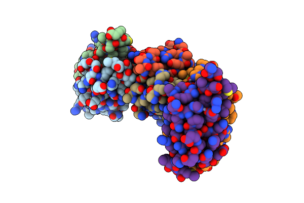
Structure Of A Native Drosophila Melanogaster Pol Ii Elongation Complex With A Well-Defined Rpb4/Rpb7 Stalk
Organism: Drosophila melanogaster
Method: ELECTRON MICROSCOPY Resolution:3.44 Å Release Date: 2026-01-07 Classification: TRANSCRIPTION Ligands: ZN |
 |
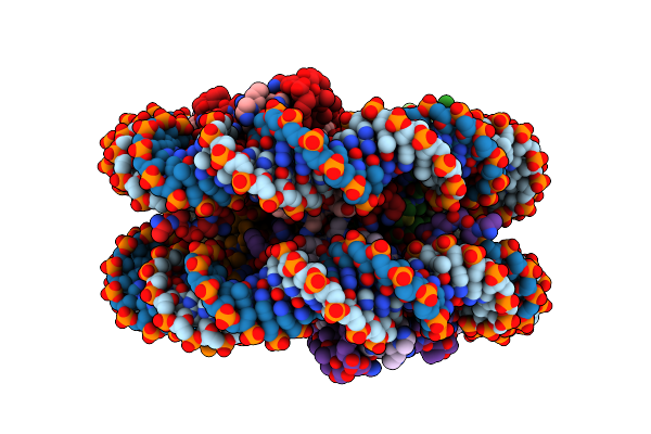
Structure Of A Native Drosophila Melanogaster Pol Ii Elongation Complex Without Rpb4/Rpb7 Stalk
Organism: Drosophila melanogaster
Method: ELECTRON MICROSCOPY Resolution:3.37 Å Release Date: 2025-05-14 Classification: TRANSCRIPTION Ligands: ZN |
 |
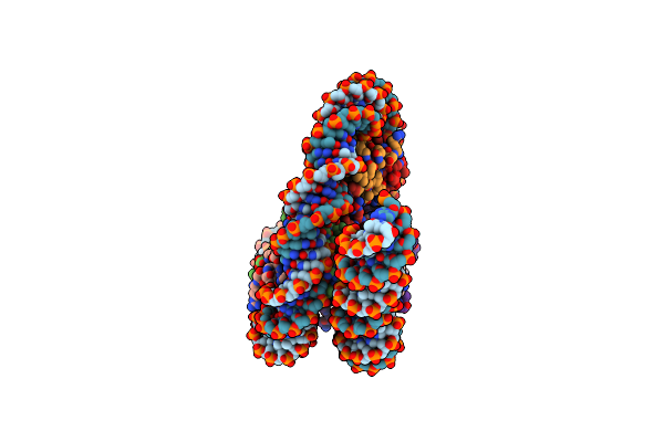
Organism: Drosophila melanogaster
Method: ELECTRON MICROSCOPY Release Date: 2025-02-19 Classification: GENE REGULATION |
 |
Organism: Drosophila melanogaster
Method: ELECTRON MICROSCOPY Release Date: 2025-02-19 Classification: GENE REGULATION |
 |
Organism: Drosophila melanogaster
Method: ELECTRON MICROSCOPY Release Date: 2025-02-19 Classification: GENE REGULATION |
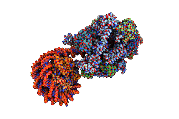 |
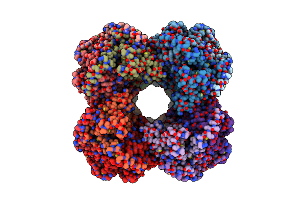
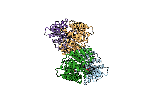
Structure Of A Native Drosophila Melanogaster Nucleosome Elongation Complex (Pol Ii Ec-Nucleosome). Composite Map
Organism: Drosophila melanogaster
Method: ELECTRON MICROSCOPY Release Date: 2025-02-19 Classification: TRANSCRIPTION Ligands: ZN |
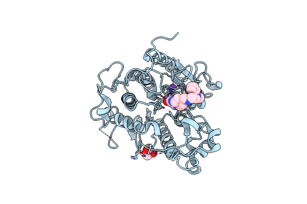 |
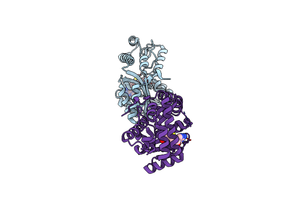
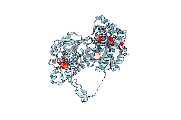
Crystal Structure Of Trypanosoma Cruzi Histone Deacetylase Dac2 Complexed With Quisinostat
Organism: Trypanosoma cruzi
Method: X-RAY DIFFRACTION Resolution:1.75 Å Release Date: 2021-12-15 Classification: HYDROLASE Ligands: ZN, K, GOK, GOL |
 |
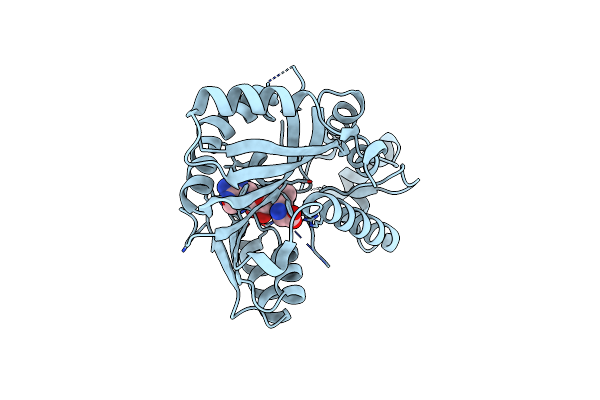
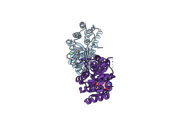
Crystal Structure Of Trypanosoma Cruzi Histone Deacetylase Dac2 Complexed With A Hydroxamate Inhibitor
Organism: Trypanosoma cruzi
Method: X-RAY DIFFRACTION Resolution:2.30 Å Release Date: 2021-12-15 Classification: HYDROLASE Ligands: ZN, K, T56 |
 |
Organism: Stigmatella aurantiaca
Method: X-RAY DIFFRACTION Resolution:1.42 Å Release Date: 2018-12-12 Classification: TRANSFERASE Ligands: SAM |
 |
Organism: Stigmatella aurantiaca
Method: X-RAY DIFFRACTION Resolution:1.96 Å Release Date: 2018-12-12 Classification: TRANSFERASE Ligands: SAH |
 |
Organism: Stigmatella aurantiaca
Method: X-RAY DIFFRACTION Resolution:1.80 Å Release Date: 2018-12-12 Classification: TRANSFERASE Ligands: SAH, CL |
 |
Organism: Stigmatella aurantiaca
Method: X-RAY DIFFRACTION Resolution:1.70 Å Release Date: 2018-12-12 Classification: TRANSFERASE Ligands: SAH |
 |
Organism: Stigmatella aurantiaca
Method: X-RAY DIFFRACTION Resolution:1.90 Å Release Date: 2018-12-12 Classification: TRANSFERASE Ligands: SAM, GOL |
 |
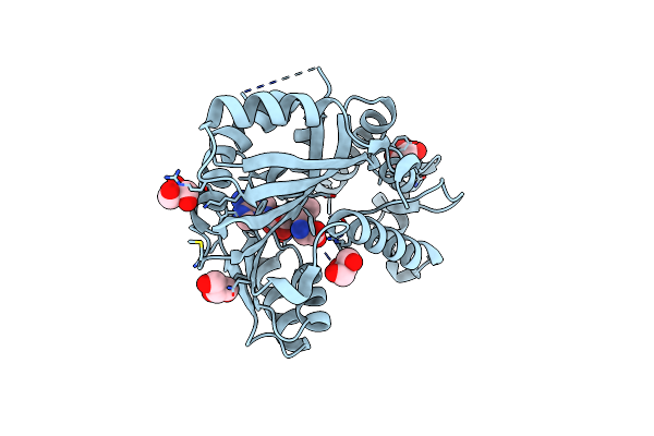
Organism: Homo sapiens
Method: X-RAY DIFFRACTION Resolution:2.51 Å Release Date: 2018-11-07 Classification: ANTITUMOR PROTEIN Ligands: DMS, F6P, BWW, ADP |
 |
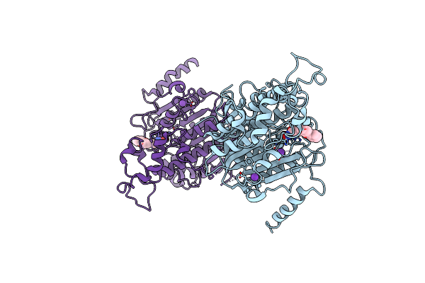
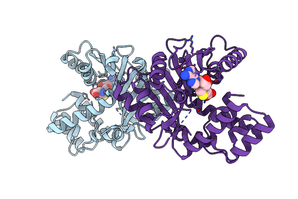
Crystal Structure Of The Murine Aum (Phosphoglycolate Phosphatase) Capping Domain As A Fusion Protein With The Catalytic Core Domain Of Murine Chronophin (Pyridoxal Phosphate Phosphatase)
Organism: Mus musculus
Method: X-RAY DIFFRACTION Resolution:2.65 Å Release Date: 2013-12-25 Classification: HYDROLASE Ligands: MG, NO3 |
 |
Organism: Archaeoglobus fulgidus (strain atcc 49558 / vc-16 / dsm 4304 / jcm 9628 / nbrc 100126), Shigella flexneri
Method: SOLUTION NMR Release Date: 2011-08-24 Classification: TRANSFERASE |
 |
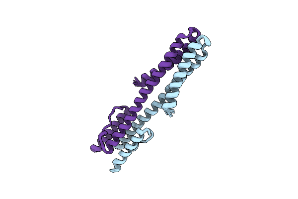
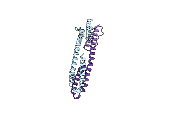
Solution Structure Of The Chimeric Af1503 Hamp- Envz Dhp Homodimer; A219F Variant
Organism: Archaeoglobus fulgidus (strain atcc 49558 / vc-16 / dsm 4304 / jcm 9628 / nbrc 100126), Shigella flexneri
Method: SOLUTION NMR Release Date: 2011-08-24 Classification: TRANSFERASE |
 |
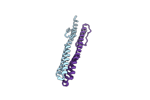
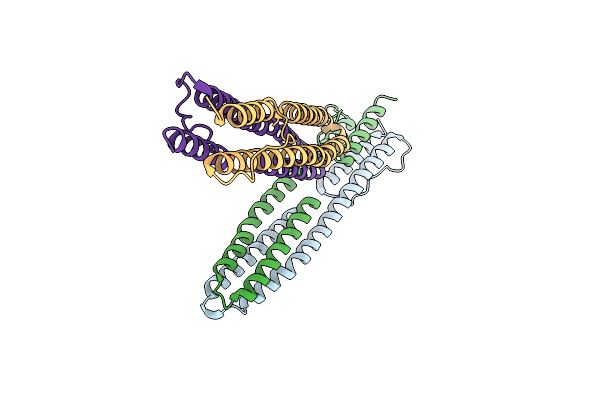
The Mechanisms Of Hamp-Mediated Signaling In Transmembrane Receptors - The A291I Mutant
Organism: Archaeoglobus fulgidus
Method: X-RAY DIFFRACTION Resolution:1.65 Å Release Date: 2011-07-20 Classification: MEMBRANE PROTEIN Ligands: ZN |
 |
The High Resolution Structure Of A Dimeric Hamp-Dhp Fusion Displays Asymmetry - A291F Mutant
Organism: Archaeoglobus fulgidus, Escherichia coli
Method: X-RAY DIFFRACTION Resolution:1.65 Å Release Date: 2011-07-06 Classification: SIGNALING PROTEIN |
 |
Organism: Archaeoglobus fulgidus, Escherichia coli
Method: X-RAY DIFFRACTION Resolution:2.25 Å Release Date: 2011-06-29 Classification: SIGNALING PROTEIN |