
Search Count: 172
 |
Organism: Homo sapiens
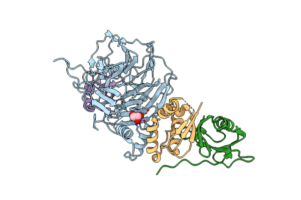
Method: ELECTRON MICROSCOPY Release Date: 2025-05-21 Classification: LIGASE Ligands: SY8 |
 |
Organism: Homo sapiens
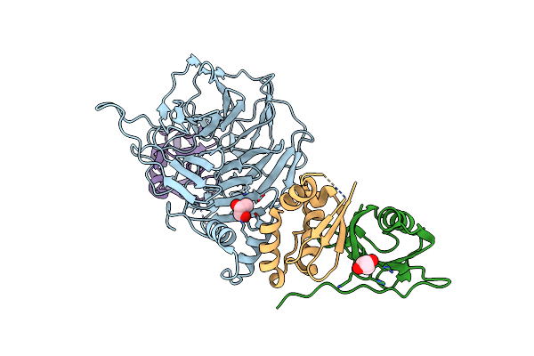
Method: ELECTRON MICROSCOPY Release Date: 2025-05-21 Classification: LIGASE Ligands: SY8 |
 |
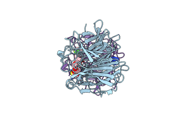
Organism: Homo sapiens
Method: X-RAY DIFFRACTION Resolution:2.00 Å Release Date: 2024-11-20 Classification: LIGASE Ligands: GOL |
 |
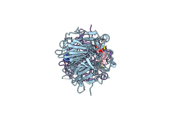
Organism: Homo sapiens
Method: X-RAY DIFFRACTION Resolution:2.60 Å Release Date: 2024-11-20 Classification: LIGASE Ligands: GOL |
 |
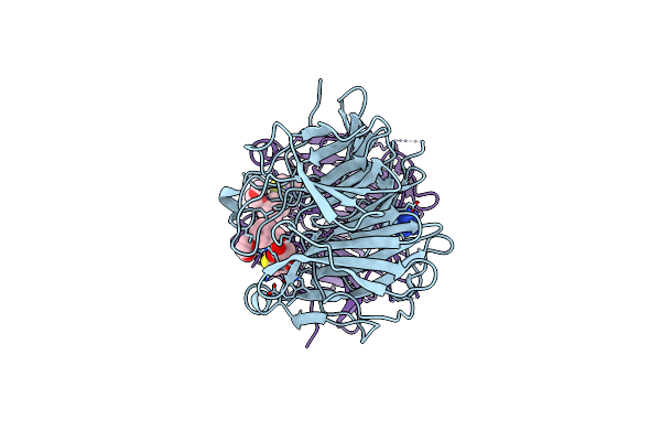
Organism: Homo sapiens
Method: X-RAY DIFFRACTION Resolution:1.88 Å Release Date: 2024-11-20 Classification: LIGASE Ligands: MPD |
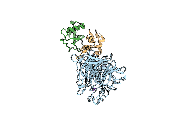 |
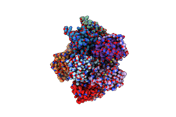
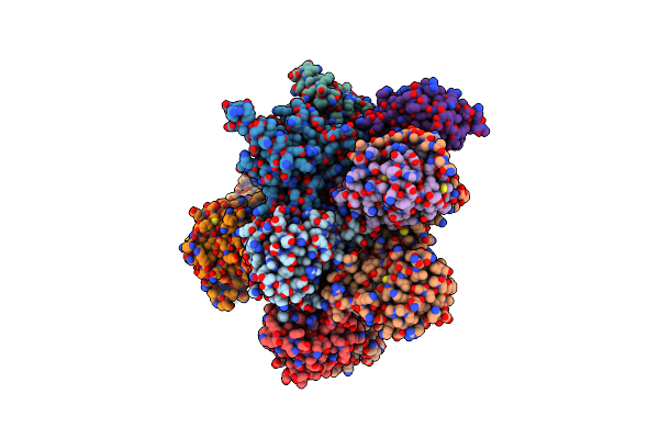
Focused Map Of Cryo-Em Structure Of Ubiquitin C-Degron Bound To Klhdc10-Elob/C
|
 |
Organism: Homo sapiens
Method: X-RAY DIFFRACTION Resolution:1.91 Å Release Date: 2024-11-06 Classification: LIGASE/INHIBITOR Ligands: A1APK, NCO |
 |
Organism: Homo sapiens
Method: X-RAY DIFFRACTION Resolution:1.70 Å Release Date: 2024-11-06 Classification: LIGASE/INHIBITOR Ligands: A1APS, NCO |
 |
Organism: Homo sapiens
Method: X-RAY DIFFRACTION Resolution:1.70 Å Release Date: 2024-11-06 Classification: LIGASE/INHIBITOR Ligands: A1APT, NCO |
 |
Structure Of The Non-Canonical Ctlh E3 Substrate Receptor Wdr26 Bound To Ypel5
Organism: Homo sapiens
Method: ELECTRON MICROSCOPY Release Date: 2024-05-15 Classification: LIGASE Ligands: ZN |
 |
Structure Of The Non-Canonical Ctlh E3 Substrate Receptor Wdr26 Bound To Nmnat1 Substrate
Organism: Homo sapiens
Method: ELECTRON MICROSCOPY Release Date: 2024-05-15 Classification: LIGASE Ligands: NMN, ZN |
 |
 |
Structure Of Cul9-Rbx1 Ubiquitin E3 Ligase Complex In Unneddylated And Neddylated Conformation - Focused Cullin Dimer
Organism: Homo sapiens
Method: ELECTRON MICROSCOPY Release Date: 2024-04-17 Classification: LIGASE Ligands: ZN |
 |
Structure Of Cul9-Rbx1 Ubiquitin E3 Ligase Complex In Unneddylated Conformation - Symmetry Expanded Unneddylated Dimer
Organism: Homo sapiens
Method: ELECTRON MICROSCOPY Release Date: 2024-04-17 Classification: LIGASE Ligands: ZN |
 |
Ubiquitin Ligation To Substrate By A Cullin-Ring E3 Ligase & Cdc34: Nedd8-Cul2-Rbx1-Elob/C-Fem1C With Trapped Ube2R2~Donor Ub-Sil1 Peptide
Organism: Homo sapiens
Method: ELECTRON MICROSCOPY Release Date: 2024-02-21 Classification: LIGASE Ligands: U9O, ZN |
 |
Organism: Homo sapiens
Method: ELECTRON MICROSCOPY Release Date: 2024-02-21 Classification: HYDROLASE |
 |
Organism: Homo sapiens
Method: ELECTRON MICROSCOPY Release Date: 2024-02-21 Classification: HYDROLASE |
 |
Organism: Homo sapiens
Method: ELECTRON MICROSCOPY Release Date: 2024-02-21 Classification: HYDROLASE |
 |
Organism: Homo sapiens
Method: ELECTRON MICROSCOPY Release Date: 2024-02-21 Classification: HYDROLASE |
 |
Organism: Homo sapiens
Method: ELECTRON MICROSCOPY Release Date: 2024-02-21 Classification: HYDROLASE |