
Search Count: 51
 |
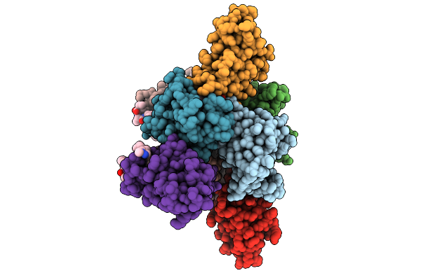
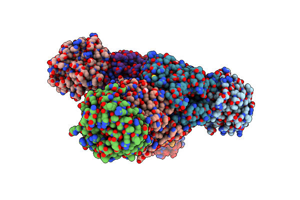
Co-Crystal Structure Of The Ternary Complex Of Human Fkbp12, Qdpr And Compound 4
Organism: Homo sapiens
Method: X-RAY DIFFRACTION Resolution:1.39 Å Release Date: 2025-10-08 Classification: LIGASE Ligands: GOL, CL, A1BB9 |
 |
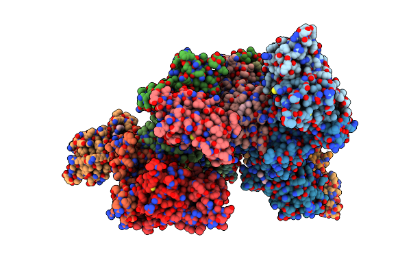
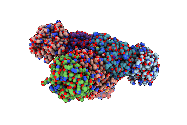
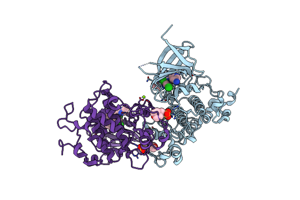
Co-Crystal Structure Of The Ternary Complex Of Human Fkbp12, Brd9 Bromo Domain And Compound 1
Organism: Homo sapiens
Method: X-RAY DIFFRACTION Resolution:2.01 Å Release Date: 2025-10-08 Classification: LIGASE Ligands: A1BB8, GOL |
 |
Dna-Encoded Library (Del)-Enabled Discovery Of Proximity Inducing Small Molecules
Organism: Homo sapiens
Method: X-RAY DIFFRACTION Resolution:3.40 Å Release Date: 2023-10-04 Classification: APOPTOSIS Ligands: X5K |
 |
Plasmodium Falciparum Cytoplasmic Phenylalanyl-Trna Synthetase In Complex With Brd7929
Organism: Plasmodium falciparum
Method: X-RAY DIFFRACTION Resolution:3.60 Å Release Date: 2022-03-23 Classification: LIGASE Ligands: B79, MG |
 |
Plasmodium Vivax Cytoplasmic Phenylalanyl-Trna Synthetase In Complex With Brd1389
Organism: Plasmodium vivax
Method: X-RAY DIFFRACTION Resolution:3.00 Å Release Date: 2020-11-04 Classification: LIGASE Ligands: FB9, MG |
 |
Crystal Structure Of Tryptophan Synthase From M. Tuberculosis - Aminoacrylate- And Gsk1-Bound Form
Organism: Mycobacterium tuberculosis (strain atcc 25618 / h37rv)
Method: X-RAY DIFFRACTION Resolution:2.41 Å Release Date: 2020-09-30 Classification: LYASE/Lyase Inhibitor Ligands: FMT, MLI, PZJ, P1T, K, EDO, PEG, ACT, SO4, NA |
 |
Crystal Structure Of Tryptophan Synthase From M. Tuberculosis - Aminoacrylate- And Gsk2-Bound Form
Organism: Mycobacterium tuberculosis (strain atcc 25618 / h37rv)
Method: X-RAY DIFFRACTION Resolution:2.40 Å Release Date: 2020-09-02 Classification: Lyase/Lyase Inhibitor Ligands: FMT, EDO, ACT, MLI, PZV, P1T, PEG, K, EPE, ALA, PGE, NA |
 |
Organism: Homo sapiens
Method: X-RAY DIFFRACTION Resolution:1.54 Å Release Date: 2020-04-01 Classification: OXIDOREDUCTASE Ligands: G9N, SO4, EDO, DMS |
 |
Organism: Homo sapiens
Method: X-RAY DIFFRACTION Resolution:1.01 Å Release Date: 2020-04-01 Classification: OXIDOREDUCTASE Ligands: CL, EOH, GOL |
 |
Crystal Structure Of Tryptophan Synthase From M. Tuberculosis - Ligand-Free Form
Organism: Mycobacterium tuberculosis (strain atcc 25618 / h37rv)
Method: X-RAY DIFFRACTION Resolution:2.46 Å Release Date: 2017-05-31 Classification: LYASE Ligands: MLI, FMT, K |
 |
Crystal Structure Of Tryptophan Synthase From M. Tuberculosis - Aminoacrylate-Bound Form
Organism: Mycobacterium tuberculosis (strain atcc 25618 / h37rv)
Method: X-RAY DIFFRACTION Resolution:2.40 Å Release Date: 2017-05-31 Classification: LYASE Ligands: MLI, FMT, P1T, CS |
 |
Crystal Structure Of Tryptophan Synthase From M. Tuberculosis - Ligand-Free Form, Trpa-G66V Mutant
Organism: Mycobacterium tuberculosis (strain atcc 25618 / h37rv)
Method: X-RAY DIFFRACTION Resolution:2.35 Å Release Date: 2017-05-31 Classification: LYASE Ligands: MLI, FMT |
 |
Crystal Structure Of Tryptophan Synthase From M. Tuberculosis - Brd4592-Bound Form
Organism: Mycobacterium tuberculosis (strain atcc 25618 / h37rv)
Method: X-RAY DIFFRACTION Resolution:2.45 Å Release Date: 2017-05-31 Classification: LYASE Ligands: MLI, FMT, 79V |
 |
Crystal Structure Of Tryptophan Synthase From M. Tuberculosis - Aminoacrylate And Brd4592-Bound Form
Organism: Mycobacterium tuberculosis (strain atcc 25618 / h37rv)
Method: X-RAY DIFFRACTION Resolution:2.40 Å Release Date: 2017-05-31 Classification: LYASE Ligands: MLI, FMT, P1T, CS, 79V |
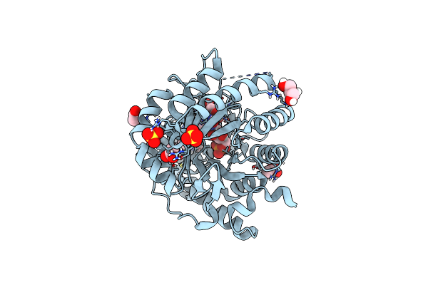 |
Organism: Homo sapiens
Method: X-RAY DIFFRACTION Resolution:1.66 Å Release Date: 2016-10-12 Classification: OXIDOREDUCTASE/OXIDOREDUCTASE INHIBITOR Ligands: FNR, ORO, MLJ, SO4, ACT, CL, GOL |
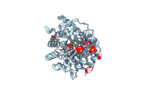 |
Crystal Structure Of Human Dihydroorotate Dehydrogenase At 1.7 A Resolution
Organism: Homo sapiens
Method: X-RAY DIFFRACTION Resolution:1.70 Å Release Date: 2016-10-12 Classification: OXIDOREDUCTASE Ligands: FNR, ORO, CE9, SO4, ACT, CL, GOL |
 |
Organism: Homo sapiens
Method: X-RAY DIFFRACTION Resolution:3.10 Å Release Date: 2016-05-25 Classification: TRANSFERASE/TRANSFERASE INHIBITOR Ligands: 65C, MG, MES |
 |
Organism: Homo sapiens
Method: X-RAY DIFFRACTION Resolution:2.45 Å Release Date: 2016-05-25 Classification: TRANSFERASE/TRANSFERASE INHIBITOR Ligands: 65A |
 |
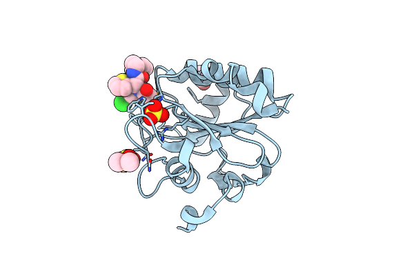
Crystal Structure Of 7,8-Diaminopelargonic Acid Synthase (Bioa) From Mycobacterium Tuberculosis, Complexed With A Thiazole Inhibitor
Organism: Mycobacterium tuberculosis
Method: X-RAY DIFFRACTION Resolution:2.24 Å Release Date: 2015-02-04 Classification: transferase/transferase inhibitor Ligands: PLP, 3GS, PEG, EDO, CL |
 |
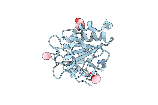
Crystal Structure Of 7,8-Diaminopelargonic Acid Synthase (Bioa) From Mycobacterium Tuberculosis, Complexed With 7-(Diethylamino)-3-(Thiophene-2-Carbonyl)-2H-Chromen-2-One
Organism: Mycobacterium tuberculosis
Method: X-RAY DIFFRACTION Resolution:1.90 Å Release Date: 2015-02-04 Classification: transferase/transferase inhibitor Ligands: PLP, 3G8, EDO, CL |